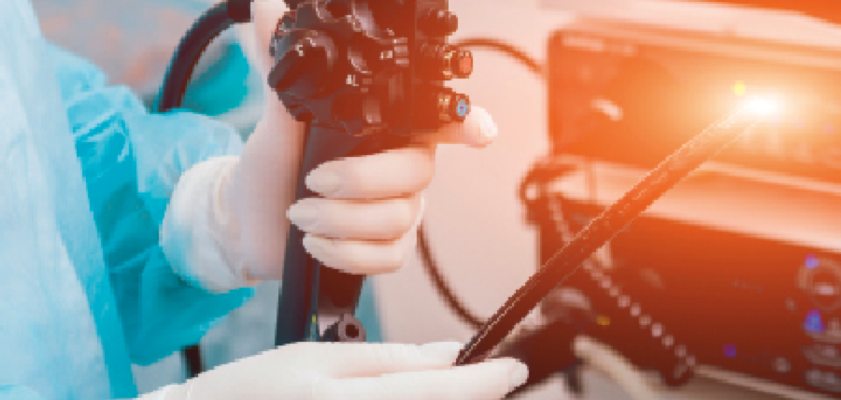
Fibre lasers are a growing presence in healthcare, with commercial products available in several applications such as vision correction, skin rejuvenation, and photo-coagulation surgery.
According to analyst IDTechEx, this is just the beginning, with technologies emerging in multiphoton microscopy, kidney stone and soft tissue ablation, and dentistry. In its latest report, Fiber Lasers 2018-2028: Technologies, Opportunities, Markets & Forecasts, the company predicts significant growth in the global fibre laser market during the next 10 years, to reach $8.9bn in 2028, with several up-and-coming technologies expected to enter healthcare in the next 10 years.
One example is bio medical imaging. This is due to the ability of fibre lasers to offer the kinds of wavelengths that ideally match novel fluorescent markers and biological molecules. Bio imaging is ideal for uses such as visualising internal organs and even diseases, with the latest technological developments claiming to offer better views of biological processes.
Live testing
Bio imaging can allow testing for the effects of various biological objects on whole, living organisms or cells and, because it is non-invasive, offers precise tracking of metabolites that can be used as biomarkers for disease identification, progress and treatment. A number of research institutes and key vendors have been working on the development and optimisation of fibre laser systems for bio medical applications.
Khalid Abou El Kabir, business development manager at Laser Components, believes that the industry has already made incredible progress. ‘The market for bio imaging has expanded over the past 10 to 20 years, so right now we are able to clearly see samples through an image. It was a tremendous improvement to get where we are right now.’
The company manufactures laser optic solutions from facilities in Germany, Canada and the US, allowing it to work alongside several research and development institutes throughout the world. El Kabir said: ‘In the UK we do not manufacture the laser, but we work with our international partners to supply lasers to customers in the UK and world-wide, who work in research facilities or universities.’
It takes two
A team of researchers at the Ecole polytechnique fédérale de Lausanne (EPFL) research institute and university, in partnership with the Department of Otolaryngology and Eaton Peabody Laboratories, Massachusetts Eye and Ear and Harvard Medical School, recently demonstrated that a multimode fibre device can provide two-photon fluorescence (TPF) imaging feedback to guide the femtosecond laser ablation (FLA) in biological samples, such as in the inner ear.
The system was implemented via the transmission of high-power femtosecond pulses through a graded-index multimode fibre that can deliver laser intensities up to 1.5 x 1,013W/cm2, which is appropriate for the ablation of biological samples. Wavefront shaping through an ultra-thin probe – around 400 micrometres in diameter – was used for diffraction limited focusing and digital scanning of the focus spot, and selective ablation of cochlear hair cells was performed based on images obtained through the same probe.
The researchers tested the performance of this dual-modality endoscopic tool in the organ for hearing (organ of Corti) from an extracted mouse cochlea. The organ includes hair cells that are responsible for the detection and transduction of sounds. TPF images of the cochlear hair cells were collected via the endoscope, so that they could be used to define the area of interest to be ablated through the same tool. By developing this technique – which integrated imaging modalities with selective laser ablation capabilities – the researchers said they have created a minimally invasive system for both cellular level investigation of distinct target areas and for cell manipulation.
Higher resolution imaging
The team – consisting of Eirini Kakkava, Marilisa Romito, Donald Conkey, Demetri Psaltis Damien Loterie, and Christophe Moser from EPFL, and Konstantina M Stankovic – Department of Otolaryngology and Eaton Peabody Laboratories, Massachusetts Eye and Ear and Harvard Medical School – reported their findings in the Optical Society of America’s research journal, Biomedical Optics Express.
Among these findings, they highlighted that the integration of TPF imaging and FLA in the same fibre in this way allows optical guided feedback that could be useful for medical investigation, diagnosis and therapy. For example, in hearing loss, the majority of which originates from the inner ear, direct visualisation of cochlear micro-anatomy would be useful to establish precise diagnosis and guide therapy, which could include morphological and functional modification of inner ear tissues.
It was also demonstrated that a device like this can perform FLA with high selectivity based on the TPF image of the sample, which is obtained through the same probe. TPF imaging provides high penetration depth for imaging of scattering samples, such as biological tissues, due to the use of infrared photons. The non-linear character of the process also results in higher resolution and sectioning than single-photon fluorescence imaging.
In addition, the cellular-level FLA selectivity demonstrated in this study could prove useful in medical applications like neurotology and neurosurgery, such as for the treatment of intracochlear or intralabyrinthine schwannomas – a rare sudden hearing loss and vertigo condition. Genomic and proteomic analyses, as well as laser microdissection of chromosomes, are additional areas where selective laser ablation could be of interest.
Commercial applications
When it comes to commercial use, this appetite for higher resolution images is a key driver for fibre laser development in medical applications, as El Kabir explained: ‘One of the big things in bio imaging is the resolution. That is why our customers try to find different techniques to get very high resolution, like sub-micro spatial resolution, and that is why they need this type of laser.’
One such technique has been developed by researchers from the Institute of Photonics Technology at Jinan University in Guangzhou, China, and looks set to have applications in wearable devices and instrumentation, as well as in diagnosis applications.
The new fibre laser-based ultrasound sensor was presented by lead researcher Long Jin at the OSA Frontiers in Optics + Laser Science APS/DLS conference last September. The technique uses optical fibre technology to provide new sensors for photoacoustic imaging, utilising fibre optic ultrasound detection, and exploiting the acoustic effects on laser pulses via the thermoelastic effect.
Jin said: ‘Conventional fibre optic sensors detect extremely weak signals by taking advantage of their high sensitivity via phase measurement.’ Sensors such as these are often used in military applications to detect low-frequency acoustic waves, but they are less effective when it comes to ultrasound waves at the kind of megahertz frequencies that medical purposes require, because ultrasound waves typically broadcast as spherical waves and have limited interaction length with optical fibres. Jin explained to delegates that the new sensors were specifically developed for medical imaging, as they can provide better sensitivity than the piezoelectric transducers in use today.
The small things
The researchers also designed an ultrasound sensor that is basically a compact laser built in the 8µm-diameter core of a single-mode optical fibre. Jin said: ‘It has a typical length of only 8mm. To build up the laser, two highly reflective grating mirrors are UV-written into the fibre core to provide optical feedback.’
It then got doped with ytterbium and erbium to provide sufficient optical gain at 1,530 nanometres, and a 980-nanometre semiconductor laser is used as the pump laser.
Yizhi Liang, assistant professor at the Institute of Photonics Technology, explained that fibre lasers such as this – which have a kilohertz-order linewidth – can be exploited as sensors due to their ability to offer a high signal-to-noise ratio. The detection benefits from the combined technique, he explained, due to the side-incident ultrasound waves deforming the fibre, modulating the lasing frequency. ‘By detecting the frequency shift,’ he said, ‘we can reconstruct the acoustic waveform.’
Mix and match
The team used a method known as self-heterodyning, in which the result of mixing two frequencies is detected. They measured the radio frequency-domain beat note provided by two orthogonal polarisation modes of the fibre cavity. This intrinsically guarantees a stable signal output.
In terms of commercial applications, the fibre sensors offer opportunities for use in photo-acoustic microscopy. The researchers used a focused 532 nanometre nanosecond pulse laser to illuminate a sample and excite ultrasound signals. They placed a sensor in a stationary position near the biological sample to detect optically induced ultrasound waves. Jin explained: ‘By raster scanning the laser spot, we can obtain a photoacoustic image of the vessels and capillaries of a mouse’s ear. This method can also be used to structurally image other tissues and functionally image oxygen distribution by using other excitation wavelengths – which takes advantage of the characteristic absorption spectra of different target tissues.’ The use of optical fibres is ideal for this, he said, due to their tiny and lightweight nature, alongside inherent flexibility.’
Jin added: ‘The development of our laser sensor is very encouraging, because of its potential for endoscopes and wearable applications. But current commercial endoscopic products are typically millimetres in dimension, which can cause pain, and they don’t work well within hollow organs with limited space.’
El Kabir believes that another key driver for the demand for fibre lasers in medical applications is their ability to very easily integrate with their systems. ‘And the beauty,’ he said, ‘is that they have a high-performance source. That is why these types of lasers contribute to this application.’
The price is right
Another attraction, as might be expected, is cost. ‘The advantage of a fibre laser is that people in the medical community like this type of laser. This is because, in terms of pricing, they have a very reasonable cost,’ he explained. ‘Secondly, because they can integrate that system very easily and also, laser vendors, such as ourselves, offer such lasers for the bio imaging community.’
As with all technological advances, there are also challenges involved when it comes to development for the commercial market. One such trial, said El Kabir, is power. ‘You have to be very cautious,’ he said, ‘because we’re talking about fibre. So, when you use a very high power, if you are not very careful about how you use the fibre, you could end up having the fibre burning, so that’s something that could be challenging for the manufacturer.’
Looking at how the industry may continue to develop commercially, El Kabir said: ‘I think it will depend on how the technology progresses, because vendors have to continue to follow up and find a better way to supply the base units for these guys.
‘For example, with imaging, there is a number of frames per second, and the higher the frame, the better the image,’ continued El Kabir.
‘Sometimes you need the laser to switch very fast, so we’re talking about frequency repetition. This depends on which direction we are going and how we can assist this community.
‘The common wavelength that these guys use is in the range of 560 to 780nm. Suppliers, such as ourselves, need to make sure that we continue to engage with them, talking with them to gauge what they are asking for and how we can best assist them,’ El Kabir concluded. EO