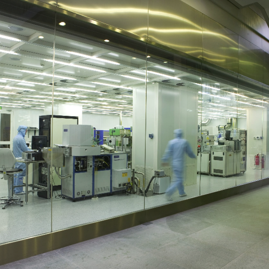
The Fraunhofer Institute for Laser Technology ILT in Aachen has developed a powerful light source for compact X-ray microscopes that will allow biological cells to be studied in high resolution. Using a technique similar to that of medical tomography, it is now possible to obtain layered three-dimensional images of biological cells or even semiconductor devices.
The task of analysing the internal structure of biological cells is a complex affair. When using an electron microscope, the whole cells first have to be fixed, followed by the time-consuming task of preparing the individual slices. The surface of the slices can then be analysed at high resolution, one slice at a time. The procedure is much less laborious when using an x-ray microscope. Immediately after cryo-fixing of the whole cells, it is possible to obtain 3D images with a resolution of 20nm (at current standards).
The technique is rather similar to that of medical tomography (CAT scanning). X-ray microscopes can also be used in semiconductor electronics to examine current-carrying circuits at high resolution. This allows defects to be detected and visualised in working electronic devices.To achieve the comparatively high resolution of 20nm that distinguishes X-ray microscopy from basic light microscopy, a short-wavelength source in the soft X-ray range is required.
Furthermore, the appropriate short exposure times call for the presence of a high photon flux. To date, the usual way of generating the necessary photon flow has involved the use of an electron storage ring. Such facilities are only available in a limited number of major research centres, and can only be used on-site, which makes it difficult for many users to take advantage of them.
The Fraunhofer Institute for Laser Technology has now developed a compact, integrated light source/collector lens system that enables powerful x-ray microscopes to be built on a laboratory scale. The volume of the resulting x-ray microscope does not exceed 2m3. This permits it to be installed wherever it is needed.
The new x-ray microscope is capable of operating with exposure times in the single-digit second range for thin samples of less than one micrometre, or several tens of seconds for larger biological samples with a thickness of a few micrometres. Dr Klaus Bergmann, who leads the Fraunhofer ILT project team, said he is certain that: 'We will be able to bring the exposure time down to below 10 seconds for the larger samples too, by optimising the design of the condenser mirror.'

