A new microscopic protein crystal selection method using laser tweezers is expected to streamline the use of x-ray crystallographic analysis of molecule structure and function.
Scientists want to study molecules to help them understand phenomena such as diseases. Developed at the Rutherford Appleton Laboratory, the new method uses a microfibre web to create a net for the micro crystals to be mounted on. The crystals can be so small, less than 10 microns, that they are difficult to handle and mount onto standard sample holders. This can lead to some potentially vital crystals being discarded.
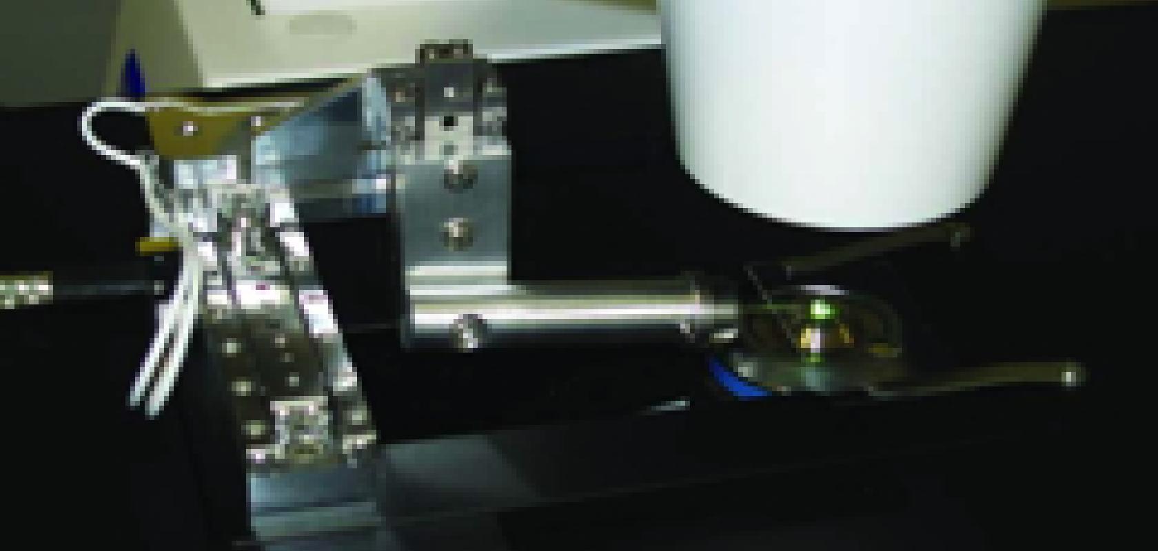
In the new method, a laser tweezer is used to move crystals onto a new type of sample holder. By using lasers to place these crystals in position, scientists are able to know where to focus the X-ray beam. The technique is the result of a cross-campus collaboration between the UK’s national synchrotron science facility, the Diamond Light Source, and the Science and Technology Facilities Council central laser facility and technology departments.
Armin Wagner, project lead at Diamond Light Source, explained why the technique is so important: 'Previously we have, in essence, wasted two to three days on the beamline just looking for our best crystals. Now, using the laser tweezers system, we can visually identify and select specific crystals and transfer them to well defined positions on a sample mount rather than relying on a purely random process. Microcrystal work is essential for research in many areas and this collaborative, cross-campus project is addressing problems with crystal supply to allow more efficient research.'
