Researchers at the University of California, Irvine are testing a new imaging device for its ability to monitor, quantify – and hopefully one day predict – skin toxicity levels induced by radiation therapy.
The team from the university’s Beckman Laser Institute (BLI) and Medical Clinic, and the Department of Radiation Oncology, used a spatial frequency domain imaging (SFDI) system developed by start-up Modulated Imaging, and presented their results at the OSA Biophotonics Congress: Optics in the Life Sciences meeting, held in April in San Diego, California.
To eradicate any cancer cells that may potentially remain after surgery or chemotherapy, many breast cancer patients also undergo radiation therapy. All patients experience side effects including skin irritation, and sometimes peeling and blistering. Patients can also develop permanent discoloration of the skin and thickening of the breast tissue months, or even years, after treatment. There is currently no method to predict the severity of these effects, and even current evaluation of these effects are based on subjective scoring.
Using the new imaging technique, the project is aimed at using precision measurements to characterise skin toxicity of tissue exposed to radiation. By tracking these measurements throughout treatment, the researchers hope to better understand the factors involved in skin damage and, hopefully, how to predict acute and late toxicities.
‘The toxicity is basically the skin damage, a side effect from the radiation,’ said Anaïs Leproux, a post-doctoral researcher at BLI. ‘We are trying to characterise the skin damage during radiation therapy, especially for the treatment of breast cancer.’
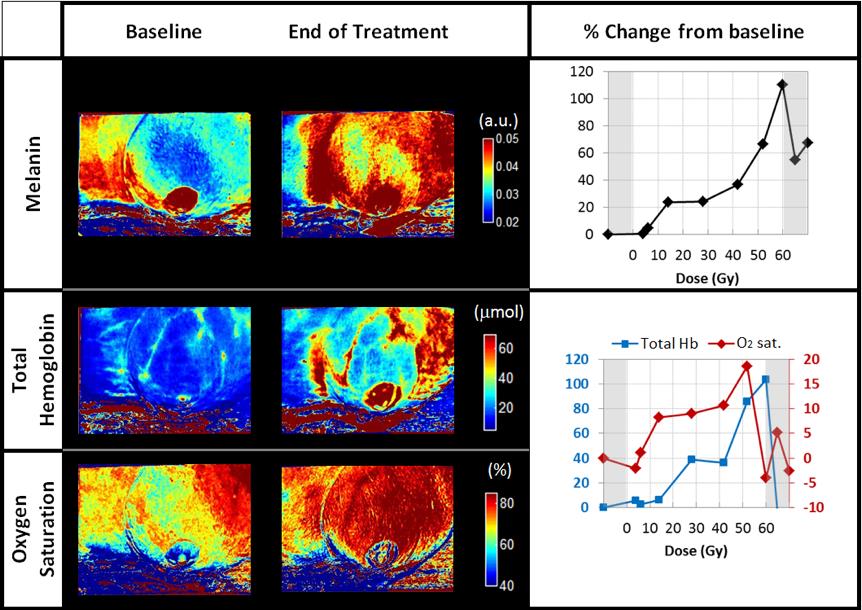
SFDI images of melanin, total hemoglobin and oxygen saturation of the treated breast of a patient, at baseline and at the end of the radiation treatment. The right panels show the per cent change from baseline of the melanin (top), and total haemoglobin and oxygen saturation (bottom) for all the study time-points. Credit: Anaïs Leproux, Beckman Laser Institute
Leproux and her group use eight different wavelengths of visible and near-infrared light from LEDs, and measure how the energy of each wavelength is absorbed by the skin. This provides them with quantitative values indicative of skin health.
To generate these values accurately, the light from the LEDs is modulated spatially, imparting distinct patterns with a digital micro-mirror device within the instrument. Formally, this functional imaging technique is called spatial frequency domain imaging, or SFDI.
‘Since we use several wavelengths of light, we perform spectroscopy and obtain the content of melanin, tissue haemoglobin, in the de-oxygenated and oxygenated state, from which we can calculate the total blood volume and oxygen saturation in the tissue,’ Leproux explained. ‘We measure superficially, about three to five millimetres deep.’
This non-invasive look at just those few millimetres can reveal a lot about the changes radiation induces. Also, because they use a projector technology, they measure over large areas (about 20 by 20cm) without scanning.
‘We’re hoping that we can see skin thickening in the scattering parameters we’re looking at,’ she said. ‘We think that the radiation induces a remodelling of the collagen in the skin, which should be seen as a change in the scattering parameter.’
Although results are still in their infancy, the successful identification of distinctly different trends in melanin and oxygen saturation demonstrates the technique’s potential.
Along with aiming to one day predict a patient’s reactions to radiation therapy, the group hopes to optimise the device in other ways along the way, perhaps helping to guide the development of better lotions to treat these side effects as well as shrinking the size of the instrument itself.
‘We could optimise the current instrument in order to have shorter measurements with a cheaper device. That’s something we’ll look into,’ said Leproux.
