Researchers from the Massachusetts General Hospital in the USA have developed an optical device that provides care-givers with timely information about the clotting properties of a patient’s blood in near real time.
The device, described in the journal Biomedical Optics Express on 24 February, is a fast and practicable alternative to current methods of monitoring defective blood coagulation - one of the major causes of preventable death in patients who have suffered trauma or undergone surgery.
The body’s natural defence against severe blood loss is the clotting process. However, in certain conditions, or when a patient undergoes surgery or suffers a trauma, blood can coagulate too little or too much.
‘Currently, the most comprehensive measures of coagulation are a battery of lab tests that are expensive and can take hours to perform,’ said Seemantini Nadkarni, an assistant professor at the Wellman Center for Photomedicine at Massachusetts General Hospital and Harvard Medical School, and senior author on the Biomedical Optics Express paper. She noted that other point-of-care systems have been developed that provide clotting measurements, but can be large, expensive, and require significant amounts of blood. ‘Our goal is to provide as much information as a lab test, but to provide it quickly and cheaply at a patient’s bedside,’ added Nadkarni.
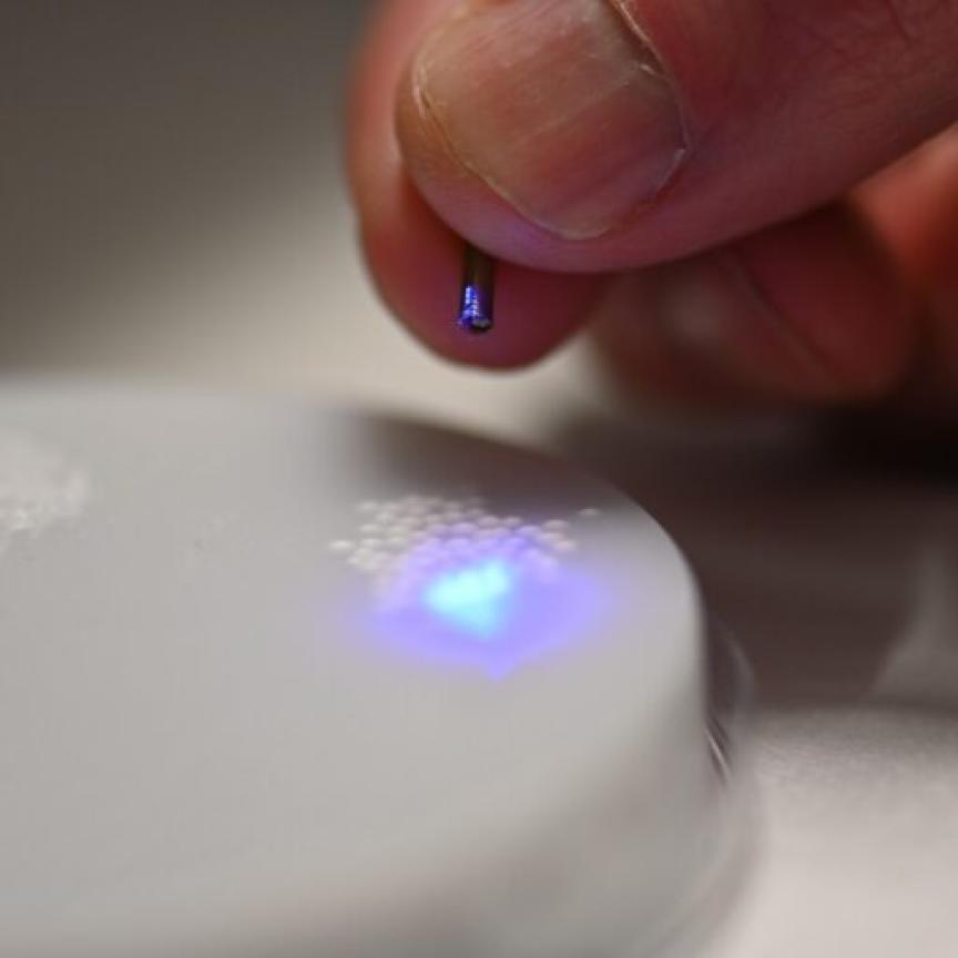
To reach this goal, Nadkarni and her colleagues turned to an optical technique they pioneered called laser speckle rheology (LSR). In LSR, researchers shine laser light into a sample and monitor the patterns of light that bounce back.
When light hits a blood sample, blood cells and platelets scatter the light. In unclotted blood these light scattering particles freely move, making a pattern of scattered light, known as a speckle pattern, which fluctuates rapidly. ‘It’s almost like looking at a starry night sky, with twinkling stars,’ Nadkarni explained. ‘But as the blood starts to coagulate, blood cells and platelets come together within a fibrin network to form a clot. The motion is restricted as the sample gets stiffer, and the twinkling of the speckle pattern is reduced significantly.’
Nadkarni and her team used a miniature high-speed camera to record the fluctuating speckle pattern and then correlated the intensity of changes in the pattern with two important blood sample measurements: clotting time and concentration of fibrinogen, a protein that plays a key role in the clotting process. Doctors in an emergency room or performing surgery could use the measurements to make decisions about how much blood to give a bleeding patient and what type of blood product, for example platelets or fibrinogen, is needed most.
‘The timely detection of clotting defects followed by the appropriate blood product transfusion is critical in managing bleeding patients,’ Nadkarni stated. ‘If you transfuse too much, there could be further coagulation defects that occur, but if you don’t transfuse enough, bleeding continues.’
On the other end of the spectrum, Nadkarni says the device could also help patients whose blood coagulates too easily, forming clots inside of blood vessels in a condition called thrombosis. These patients take anticoagulation medications and must regularly visit labs to have their blood analysed and the doses of the medications adjusted. By having a small device that could take the same measurements in a doctor’s office or at home, could reduce the cost and inconvenience while increasing the safety of anticoagulation treatment.
Currently the optical device developed by Nadkarni and her colleagues is about the size of a tissue box and is connected to a computer. The team is working to further miniaturise the system and aims to perform clinical studies with a handheld version smaller than a cell phone within the next year.
‘I look forward to working on the exciting next phase in which we plan to conduct clinical testing of the LSR device at the point of care in the operating room and in the doctor’s office using just a drop or two of blood,’ said Markandey Tripathi, a postdoctoral fellow at the Wellman Center and lead author on the Biomedical Optics Express paper.
‘Some other rapid devices exist but these have various disadvantages, ranging from poor correlation with central laboratory tests to skill required to interpret results,’ added Elizabeth van Cott, who is an associate professor of Pathology at Massachusetts General Hospital and Harvard Medical School, and a co-author on the paper. ‘The capability of the LSR device to provide rapid test results using small amounts of blood would be extremely valuable for patients particularly in operating suites, emergency departments, and intensive care units, as well as for any patient with a coagulation disorder.’