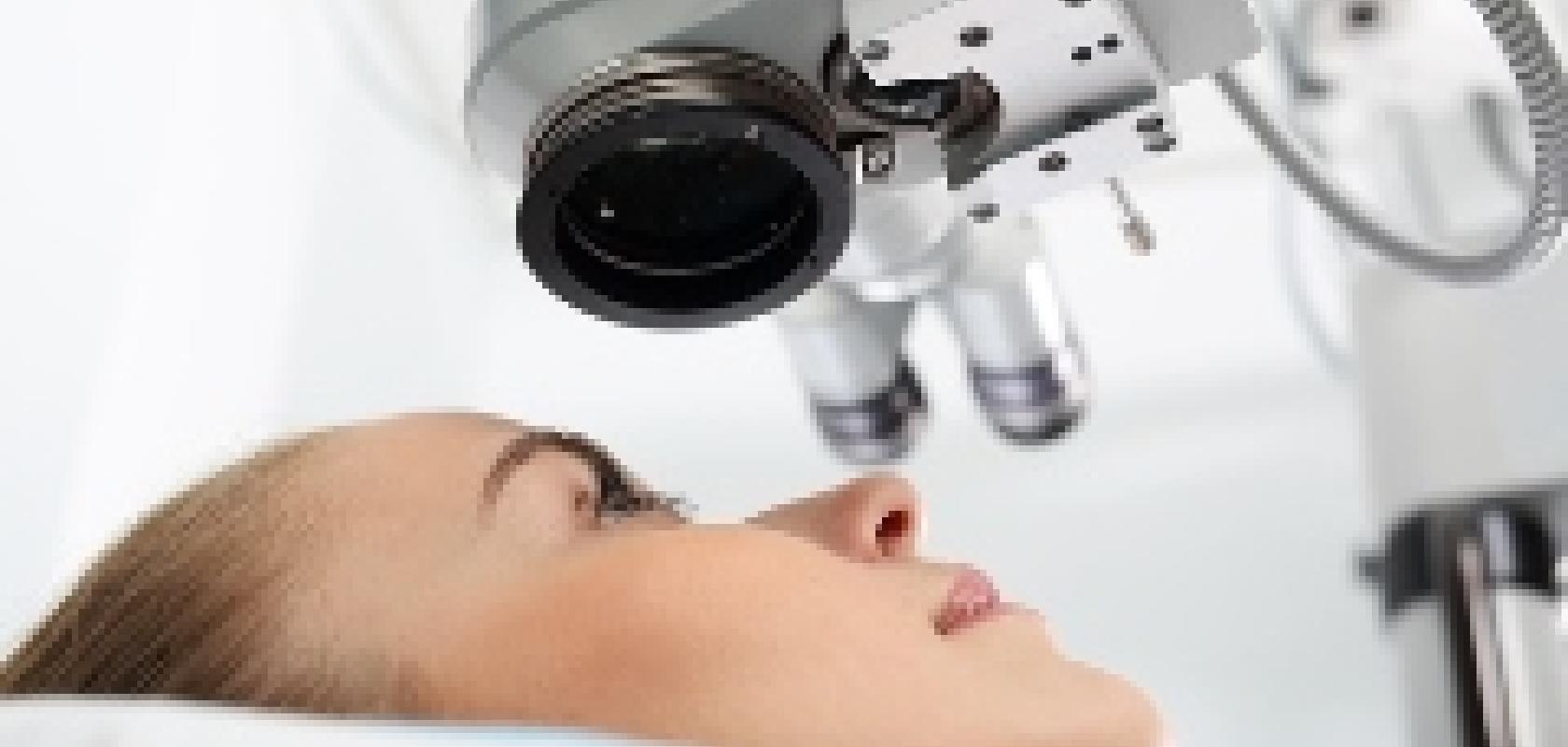
German scientists have combined femtosecond lasers with optical coherence tomography (OCT) in order to treat visual defects which cannot be corrected through existing laser-based treatments.
The new technique is a result of a project called ‘Innovative cataract, presbyopia and retinal treatment using ultrashort laser pulses’ (IKARUS), which involved research institute for photonics and laser technology, Laser Zentrum Hannover (LZH), and four industrial project partners.
Presbyopia is a common type of vision disorder that occurs as a person ages, resulting in the inability to focus up close.
As a person gets older, the central gel that fills the eye – vitreous – liquefies and loses shape, leading to separation of the vitreous from the retina located at the back of the eye. This separation is a normal part of aging and is called a posterior vitreous detachment (PVD). However, if the separation is not complete, small areas of the vitreous can remain attached to the macula, the part of the retina that is responsible for sharp, central vision, which results in distorted vision.
Currently, the most common laser surgery for correcting short-sightedness is laser in-situ keratomileusis (LASIK), whereby the cornea is cut open using a femtosecond laser to subsequently correct the defective vision.
In order to treat presbyopia however, the tissue has to be cut deeper. The scientists at the LZH and their industrial partners use a femtosecond laser for precisely cutting the lens, creating slip planes and thus making the lens more flexible.
In order to remove retina adhesions, currently the eye must be opened and the vitreous body removed. The scientists at the LZH have integrated adaptive optics into a femtosecond laser system, to be able to cut close to the retina. First results have already been established on the retina of pig eyes: with this system, model membranes only a few hundred micrometres away from the retina have been cut, with the retina tissue directly behind it showing no noticeable damage.
This treatment is only possible through an effective visualisation of the eye tissue. For this, the Image Guided Laser Surgery Group of the Biomedical Optics Department of the LZH has adapted an OCT imaging unit from German femtosecond laser company Rowiak. Using this system along with software it is possible to image the cutting of the eye as well as the laser beam delivery during the operation. Within the project, cuts into the eye have already been made without damaging either the front or the rear part of the lens capsule. Rowiak is further examining this process in ongoing clinical trials.
Apart from the LZH and Rowiak as the system manufacturer, other industry partners for the IKARus project include: Qioptic, KG (Göttingen) as optics designers, Arges (Wackersdorf) for laser scanner systems, and the Laserforum Köln (Cologne) for clinical consulting and for measuring of the eyes.
Related stories
MIT eye scanner to aid early detection of retinal diseases
Further information


