Testing beauty products to determine if they can in fact hydrate the skin or smooth fine lines not only helps to justify those high prices, but it is becoming increasingly important in brands’ marketing strategies.
The cosmetics industry relies on a variety of light-based techniques to ensure that its products are safe, effective and live up to promises made on the packaging.
And, because companies want to obtain ever more detailed information on the efficacy of their products, there is a constant demand for analytical techniques that can measure further beneath the surface of the skin in a non-invasive and non-destructive way.
‘The cosmetics industry is always hungry for new technology that can demonstrate the activity of their products. It is sometimes the same activity, but cosmetics companies are always looking for new ways to demonstrate it,’ said Laurent Peno-Mazzarino, technical director at BIO-EC Laboratory. ‘Cosmetics companies understand that marketing triggers the buy, but research leads to efficacy and triggers the rebuy. So, it is very important to invest in real research to make the best products.’
Based in France, BIO-EC Laboratory conducts research for various cosmetics enterprises, from raw material providers to companies selling finished beauty products to instrument manufacturers.
Peno-Mazzarino noted that the affluence of this industry means that many advanced techniques are used as standard in cosmetics labs; electron microscopes, spectrophotometers, bioluminescence measurements, confocal microscopes, and 3D scanners are commonplace, he said.
However, despite the plethora of technolgies available, there is still a need for accurate, robust systems for deep, non-invasive imaging.
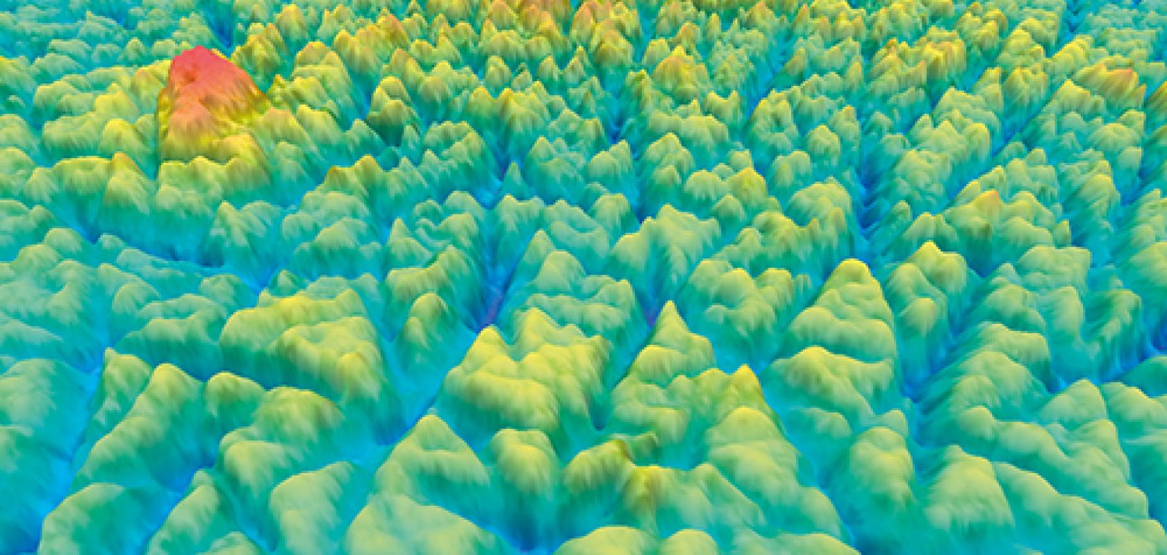
3D skin surface topography carried out by the BIO-EC Laboratory

Skin deep
BIO-EC Laboratory was founded in 1998 as a result of a research project that developed a human ex-vivo model; a piece of skin, around 1cm2, which can be used to analyse the effects of cosmetics and dermatology products. ‘It’s comparable to real skin conditions; so, it’s close to in vivo evaluation on volunteers,’ said Peno-Mazzarino.
This skin model acts as an intermediate stage between cell culture and in-vivo analysis, Peno-Mazzarino added. ‘Cell culture is used for screening raw materials, but we cannot put finished products on cell cultures. The other option is clinical evaluation on volunteers, but the investigation is restricted to non-invasive and non-destructive techniques,’ he explained. ‘[The ex-vivo model] is a complementary step.’
Numerous factors can be investigated for cosmetics, not least of all skin hydration, ageing, penetration of applied molecules, analysis of residual compounds, and skin typing. Raman spectroscopy is one technique cosmetics researchers are using to evaluate the permeation of beauty products through the skin layers. Infrared light is passed through the skin and, analysing the scattered spectra of this light, the condition and quantity of substances inside the skin can be determined. ‘In combination with mapping or imaging, it is possible to generate images based on the sample’s Raman spectrum,’ said Dr Mathieu Boiret, applications manager at Horiba Scientific, which supplies a range of analytical systems for cosmetics research.
Raman spectroscopy uses near-infrared light to examine skin in-vivo, in its natural condition. ‘Raman spectroscopy is well-adapted for [cosmetics research] as it allows fast measurements, without destruction of the sample and without using any solvents,’ Boiret added.
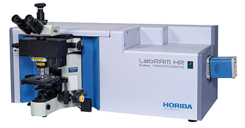
Horiba Scientific's Raman instrument, the LabRam HR Evolution, is used for cosmetics research
Last year, a study1 conducted by researchers from the University of Reims Champagne Ardenne in France reviewed Raman spectroscopy as a tool for analysing how products travel through the skin. The study concluded that Raman is an ‘analytical, non-destructive, and dynamic method to evaluate the permeation of actives in the skin layers’. The team used Raman micro-imaging to monitor the skin penetration of hyaluronic acid (HA) of different molecular weights, and found that HA with low molecular weight passed through the skin more effectively than high molecular weight HA.
Nanoscale topography is another technique used to determine how products react with the skin, but this time on the surface. Although not new, the method has only recently entered into cosmetics labs, according to Peno-Mazzarino. ‘This is a really new use of this technique,’ he said. Nanoscale topography is usually employed for surface characterisation of precision mechanical components.
BIO-EC uses a white light interferometer from Polytec to conduct surveys of the topography of the skin. A light beam is reflected by skin’s surface, and the interference of the light is measured according to a reference light beam, and a 3D representation of the top of the skin is generated.
‘The surface of the skin is composed of many micro-folds, and this micro-relief of the skin is largely responsible for the optical properties of the skin. Therefore, the image differs with the changing depth and width of the folds,’ Peno-Mazzarino explained.
Many cosmetic products try to modify the surface aspect of the skin, Peno-Mazzarino added, such as ‘soft focus’ or ‘filling’ creams that aim to reduce the depth of the micro-relief and make the skin smoother.
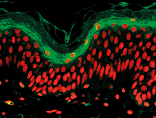
Immunostaining of LEKTI in fluorescence microscopy carried out by BIO-EC
‘We take measurements before the product has been applied, and then anywhere between five minutes and a few days after application. We can then compare the roughness of the skin and the width and depth of the micro-folds before and after treatment,’ said Peno-Mazzarino. With the ex-vivo model, the experiment can be maintained for 10 to 12 days.
These types of products that target the appearance of the skin’s surface tend to have much more immediate effects, which represents a new type of development of cosmetics, Peno-Mazzarino added.
Nanoscale topography and Raman are just two of many techniques used by BIO-EC and other cosmetics labs to provide companies with accurate information about the safety and efficacy of their products. However, when it comes to testing on human volunteers it becomes more challenging, as this requires tests that are not only minimally invasive, but robust.
‘Many existing techniques are not suitable for use on volunteers because they are too invasive or damaging. We have a lot of techniques that could be very accurate and can go deep, but are destructive. Raman spectroscopy or IR spectroscopy can sometimes be destructive or invasive. Micro-tomography x-ray… is very accurate; it can go very deep into the skin, but we cannot expose the volunteers to x-ray frequently, it is just not possible,’ Peno-Mazzarino said.
‘The light interferometer… is very sensitive to external vibrations, so it cannot be used in clinical evaluation on volunteers. We cannot put the arm or the face under the light of the interferometer because there are a lot of vibrations. If a light interferometer could be used on volunteers it could be a real advance because there is a lack of techniques for clinical evaluation,’ he continued.
‘So if there were to be an accurate, non-invasive technique that allows you to go deep within the skin it could be a real advance.’
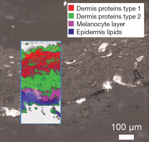
High resolution multivariate image of a skin cross section showing different constituents
Destructive but effective
When being invasive doesn’t matter, more aggressive techniques can be used to gain even more useful insights into the performance of cosmetics.
As part of a collaboration between several research and commercial organisations in France, state-of-the-art equipment and services are being made available to companies in a bid to structure research and support France’s status as leaders in cosmetics development.
The Cosmetomics innovation platform involves partners such as BIO-EC, Polytec, the University of Cergy-Pontoise, ISIPCA (a school for post-graduate studies in perfume and cosmetics products) and Soleil, a synchrotron facility near Paris.
With the particle accelerometer provided by Soleil, x-ray micro-tomography can be carried out on a hair follicle, to measure all of the components of the hair, such as the sebum-producing, or sebaceous, glands.
‘It is useful because a lot of cosmetics products aim to reduce the amount of sebum produced. With x-ray micro-tomography we can measure the volume of this gland… it is very invasive, but very useful,’ said Peno-Mazzarino.
‘A lot of big companies use these kinds of technologies. But the Cosmetomics platform allows smaller companies and research labs to access this equipment. ‘I think this is the future of research in cosmetics because a lot of these technologies cannot be acquired by a standard research lab,’ he concluded.
