At this year’s Photonics West in San Francisco, the French competitiveness cluster Route des Lasers will represent a number of photonics companies situated in the Bordeaux region. While not exhibiting at the show, the latest startup to join the cluster is Poietis, a 3D printing company. However, rather than printing inanimate objects, its technology uses a laser to print live cells.
Poietis was spun out of the French National Institute of Health and Medical Research (Inserm), based at the University of Bordeaux, late last year. Its bioprinting technology is the climax of 10 years worth of research.
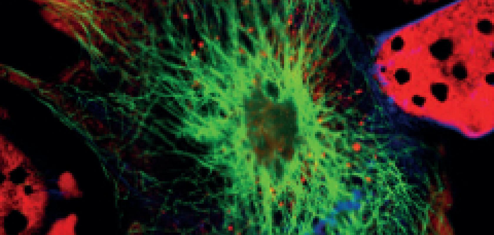
Fabien Guillemot, CEO of Poietis, said that this is one of the first companies to use lasers to 3D print living tissue and that the team is currently working on producing skin samples for pharmacological applications. He explained that this is a big growth area, as new legislation from the EU is limiting animal testing in the cosmetics industry. There is therefore a demand for affordable alternatives that could be used.
When the company was founded, ‘the aim was to develop a technique to deposit biological compounds onto biological materials’, said Guillemot. The team focus the laser onto a glass slide which is covered in a liquid film of living cells, and neighbouring a growth substrate. By focusing the light at the interface between the glass slide and the liquid film, the system induces a cavity that expands to push the liquid into a droplet. This droplet is used to deposit the live cells onto the substrate.
Bertrand Viellerobe, CTO at Poietis, explained: ‘Today, the main thing is to create this cavity which will give the capability of producing the jet. Considering the laser interaction with either the glass or the cell-carrying liquid, we want to be able to produce the cavity mentioned before but while producing very low thermal effects; we really don’t want to cook the cells. This is why we use short pulses because the thermal effects are lower.
‘To get this cavity at the right size within the right timeline, you need to use a laser with short pulses, sufficient levels of energy, and with a repetition rate compatible with the number of droplets you would like to print per second.’
In the future, Poietis will try to print other tissues such as the liver, or micro-livers, which could be used for pharmacological purposes, such as drugs testing. The company is also looking at producing corneas for the eye and also hopefully bone. ‘One of the main reasons we founded Poietis is to make the technology available for clinics,’ said Guillemot. ‘Today we focus on industrial applications, but within five or ten years, we aim to have clinical applications available.’
As of yet the system is not robust enough and there is not enough known about the cell patterns required for a tissue for it to be used in clinical applications.
‘When we consider how a tissue is formed in the body, there is a strong relation between form and function. That is to say, the positioning of the cells is very important to the function of the tissue,’ commented Guillemot. He gave the example that when experimenting with printed bone tissue in mice, the team found that different cell structures produced different bone healing profiles. So, by controlling the positions of the cells at the microscale accurately, the way in which cells interact with each other and how the tissue acts on the macroscale can be controlled.
Because of the strong link between cell patterns and tissue function, being able to guarantee each cell is where it needs to be is crucial. Therefore the resolution and accuracy of a bioprinting system is important.
Laser pulses, as opposed to mechanical methods, produce the best printing resolution, Guillemot said. The droplet volume is in the range of picolitres and the droplet diameter is 10-20µm, so the resolution of the machine is compatible with printing a single cell. Other techniques have a resolution of a few hundred microns, typically 500µm according to Guillemot.
The other big benefit of this method is the viability of the cells. The cavity formed by the laser pulse is less damaging to the cells than the mechanical equivalents, according to Viellerobe. ‘Our method allows us to print single cell resolution with very high viability, nearly 100 per cent, of the cells,’ he said.
Cell care
Cell viability is a recurring problem across the field of biophotonics. Unfortunately, using light to observe a cell can damage it – an occurrence known as photo-toxicity – which can be particularly problematic when trying to image live specimens. Jim Owens, systems sales manager at Hamamatsu Photonics UK, explained the problem: ‘Fluorescence imaging is an invasive technique whereby the light intensity required to generate a suitable signal from live cells or tissue degrades the sample. Ideally researchers would like to limit this photo-toxicity by using lower light intensities that are directed only to the cellular region of interest for the minimum amount of time.’
By having a more sensitive detector this problem can be reduced. Shorter exposure times and lower laser power intensity can be used, because the camera can capture the sample signal more efficiently, thus reducing the light-induced damage.
Owens stated that this is one of the reasons why uptake of scientific CMOS (sCMOS) sensors is growing. ‘SCMOS differs from the CCD sensors used previously in that they can deliver research-grade images in very low light-level scenarios at very high speed. Traditionally, some trade-off was required between resolution, sensitivity and speed for CCD-based cameras used in scientific research, but with sCMOS cameras such as the Orca Flash4.0, it is now possible to perform high speed, high resolution imaging at even the lowest fluorescence signal intensities.
‘It’s a really exciting, fast-moving field at the moment and advances in sensors and probes have enabled techniques that circumvent the resolution limit of light to see structures in finer detail than ever before,’ he continued. He is referring to the 2014 Nobel Prize in Chemistry, which went jointly to Eric Betzig, Stefan Hell and William Moerner for the development of super-resolved fluorescence microscopy.
Owens said that because of the award, super-resolution imaging has grabbed a lot of headlines. He explained that the process requires the researchers to take a large number of images and selectively have different parts of the sample emitting light. ‘By combining many thousands of these images, you end up with a very clear and high resolution image of the sample you are observing. The whole process has been speeded up by these new sCMOS sensors as they can take many images very quickly, meaning that it is now possible to see protein dynamics in real time.’
These new approaches are beginning to be commercialised, with Stimulated Emission Depletion (STED) microscopes and super-resolution (STORM, PALM, etc) microscopes available from the major microscope manufacturers. Owens pointed to light-sheet fluorescence microscopy as the next area that is generating considerable interest.
This is a technique that counters some of the photo-toxicity concerns mentioned, meaning that it allows for less intrusive imaging of biological samples in 3D over many hours. By subjecting the sample to illumination by a thin sheet of light, only the molecules near the focal plane are excited. This optical-sectioning results in less photo-toxicity and consequently the cells can be imaged over extended periods. Owens explained that this holds particular appeal for imaging of large organisms such as embryo development.
Hamamatsu developed a patent-pending light-sheet mode in its Orca Flash4.0 sCMOS camera as a result of customer feedback on how the sensor’s rolling shutter could be used to create a confocal slit detector when synchronised with the scanned light-sheet illumination beam. Owens said: ‘This is a good example of how being close to our customers informed our camera development for this fast moving application area, allowing us to contribute another tool for pioneers and commercial partners to incorporate into their microscope setup.
‘Light-sheet microscopy is attracting a great deal of attention from the research community and early commercial products from Zeiss, LaVision BioTec and Applied Scientific Instrumentation, as well as the Open SPIM open access platform are already available,’ he added.
Better together
When designing new products to serve new markets, such as what will have to be done to commercialise the light-sheet approach, close collaboration with customers is essential, and Owens noted there has been an expansion in life science research teams’ expertise over the past decade. ‘People used to be either a physicist or a biologist or a chemist, but now things like biophysics or biochemistry are huge growth areas,’ he said. ‘We are noticing a lot of cross-discipline research groups containing biologists, physicists and chemists as well as computer scientists and mathematicians, meaning a whole host of different expertise in one team. This has meant commercial companies can work a lot more closely with customers having a far greater spread of domain knowledge, and they have a much better understanding of what we do, while we have a better insight into the biophotonics challenges they face.’
The development stage is essential to make sure a product meets the customer’s requirements. Gooch & Housego’s marketing and communications manager, Adrian Chance, said: ‘The majority of our activity in this area is commercially orientated.
‘We are working with the leading manufacturers of ophthalmic systems already and they often have experts of photonics in-house, and they normally know, at least in theory, what they need. But when it comes to turning this into a reality, we know how to cater for those needs and the limits of what can be manufactured. Our expertise in photonics means that we can apply the technology to the customer’s needs.’
At the moment, biophotonics is not regarded as G&H’s largest sector, but in the medium to long term it is seen as a large area of growth. In the next couple of weeks the company aims to launch a biomedical imaging section within its website, in order to better serve the biophotonics market. The company’s Torquay based Systems Technology Group (STG) was set up, partly with the aim of boosting G&H’s activity within the market. ‘The STG growth was given a significant boost by European Regional Growth Funding last year, and one of their key areas is biomedical imaging,’ said Chance.
Regarding market competition, Chance explained that some biophotonic technologies are maturing and some of the leading OCT companies observed less growth than expected simply because there is more completion within the marketplace. ‘As the technology becomes established, there will always be companies producing cheaper imitations and equivalents.’
 Tom Eddershaw is a technical writer for Electro Optics, Imaging & Machine Vision Europe, and Laser Systems Europe.
You can contact him on tom.eddershaw@europascience.com or on +44 (0) 1223 275 478.
Find us on Twitter at @ElectroOptics, @IMVEurope, @LaserSystemsMag and @ESTomEddershaw.
