The contemporary phenomenon of ‘selfie’, self-taken, pictures wouldn’t be anywhere near as popular if taking an image caused the subject to disintegrate. But that’s just what happens in super-resolution fluorescence microscopy techniques that can follow nanoscale processes inside living cells.
Super-resolution microscopy’s achievements in pushing the resolution of imaging living cells below Abbe’s diffraction limit of 200nm are remarkable. But to fully exploit its capabilities, a single still snapshot of a biological system isn’t enough – changes must be monitored over time. And this is a challenge, either because processes happen too fast, or the intense light used for imaging progressively disturbs them and even kills the cells.
Yet the technology’s promise is so great that scientists in academia and industry are working to overcome this obstacle, and in doing so are gaining even more important biological insights.
Stimulated emission depletion (STED) microscopy is the fastest established technique for imaging beyond the diffraction limit. Before performing STED or any other super-resolution technique, structures of interest must be given fluorescent labels – a difficult scientific exercise in itself. STED then uses two laser sources, one focused beam that excites the fluorophores, and a second doughnut-shaped depletion beam quenching the fluorescence at the edges of that spot. Scanning the beams across a sample builds up a high-resolution picture pixel by nanometre-scale pixel. However, the laser power needed bleaches the fluorophore and damages cells, hampering long-term monitoring and other in-depth studies.
Leica Microsystems in Mannheim, Germany, supplies STED instruments, with ‘much more than 100’ operating in the field, according to Jochen Sieber, Leica’s product manager in super-resolution technologies. To avoid phototoxicity, Sieber noted that it’s important to use as many photons obtained from the sample as possible, and excite fluorophores at the optimal wavelength.
Leica designed the TCS SP8 STED 3X product specifically to achieve these aims. ‘The high sensitivity and low noise Leica Hybrid detector along with the spectral detector maximises the photons captured,’ Sieber said. ‘The Leica White Light Laser enables users to set the excitation exactly to the maximum of the excitation spectrum, be it for green, yellow or red fluorescent protein, for example.’
Sieber also pointed out that in many cases STED can provide adequate results without exploiting its full signal-to-noise or frame rate potential. ‘One should of course always be aware of the minimum requirements needed to answer a given question and design the experiment from there,’ he said. ‘What is the frame rate I need to capture the biological processes I am interested in? If you are able to go with half the speed, you’ve already halved the potential phototoxic effects.’
PicoQuant, of Berlin, Germany, also supplies a STED-capable instrument, the MicroTime 200 STED, which is highly specialised and designed to specifically perform single-molecule experiments with a super-resolution option. Called EasySTED, the approach was originally developed by 2014 Nobel Prize winner and STED inventor Stefan Hell, along with colleagues at the German Cancer Research Centre in Heidelberg, Germany1.
‘We couple the excitation laser and the STED laser into and out of one fibre – two perfectly overlaid beams,’ explained Olaf Schulz, the company’s microscopy sales specialist. ‘The EasySTED principle is to have a phase plate just before the objective that progressively changes the STED laser beam’s polarisation, but leaves the excitation laser beam unaffected. Then, interference in the focal plane gives the doughnut, and that’s really extremely robust. One time, we had an unexpected visit from a customer who wanted to see STED, so the microscope was not prepared beforehand – the phase plate was still lying in its case. So I took it out, put it into the microscope and we got sub-50nm resolution.’
One consequence of this design is that the phase plate can only be used in the 595-660nm wavelength range. This rules out standard green and yellow protein fluorophores; however, the lower-energy, red photons are less phototoxic, penetrate deeper into cells, and scatter less. In addition to providing STED super-resolution via EasySTED, the MicroTime 200 platform also integrates an atomic force microscope and a spectrograph.
The MicroTime 200 instrument is designed to have high sensitivity that allows users to turn down laser power to help minimise damage. It also provides the option of shifting the delay between excitation and STED laser beams to reduce fluorophore bleaching. ‘This accounts for fluorescence lifetimes of different labels,’ Schulz said. ‘At a specific delay you get the resolution while minimising bleaching.’
Sheet feat
Limitations still frustrate biologists studying phenomena like cell division, according to Dr Wesley Legant from the Janelia Research Campus of the Howard Hughes Medical Institute in Ashburn, Virginia.
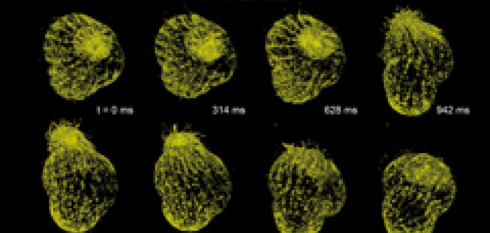
Cell division happens quickly in three dimensions, and so requires intensive laser scanning by STED, but the process is particularly sensitive to such investigation. ‘When you start trying to do things like STED, you have to illuminate the sample multiple times to get a single three dimensional image,’ Legant commented.
Even lower-intensity structured illumination microscopy (SIM), which involves shining patterned light at a sample and obtaining super-resolution information from interference arising from the resulting interaction, faces this issue. ‘The main problem is that you’re illuminating the entire 3D depth of the specimen, but only detect a single 2D plane that’s in focus. To do 3D imaging, the sample is completely exposed to illumination light for the entire duration, as you’re stepping the focal plane through the sample,’ Legant added. ‘And regardless of the technique, if you need high or deep resolution then you need to acquire more images per sample at that resolution. The need to acquire more images leads to potentially higher phototoxicity.’
Consequently, Legant and his colleagues, including Janelia Research Campus’ Dr Eric Betzig, (who won the 2014 Nobel Prize together with Stefan Hell and Stanford University’s W.E. Moerner), have been exploring an illumination scheme known as lattice light sheet microscopy.2 ‘Light sheet microscopy is a general term for bringing in illumination at 90° to the focal point,’ Legant said. ‘You’re only illuminating the single focus, and therefore the amount of light that’s going through is much less. That allows [for] improvements in terms of phototoxicity.
‘It’s also more sensitive than most other approaches. The number of volumes and the speed at which those volumes can be acquired is significantly faster than you can get out of point scanning or confocal microscopes, at a resolution that is comparable.’
While Legant, Betzig and colleagues published work combining light sheet illumination with SIM in 2014, he stressed that it could potentially be applied to any super-resolution microscopy technique.
Legant also emphasised the importance of optimising fluorophores, including the crucial capabilities of permeating into cells and directing them to their destination. Legant works closely with Dr Luke Lavis from the Janelia Research Campus, who has recently developed new types of dyes that ‘tend to be a lot brighter and a lot more photostable’.
Leica’s Sieber echoed this point, saying that improved fluorophore and labelling technologies, ‘have the same potential to bring the field forward as has instrument design’, he noted. ‘A nice example is the silicon-rhodamine (SiR) dyes for live cell STED imaging in the deep red range.’
For live cell imaging, fluorophores often feature chemical structures that selectively associate with proteins like SNAP-tag introduced to cells by genetic modification. Such tagging is limited to cells and organisms that can easily be genetically modified. However, Professor Kai Johnsson’s École Polytechnique Fédérale de Lausanne, Switzerland, team, which developed the SiR dyes together with Hell, also conjugated them to tags for cell cytoskeleton proteins3.
The developers showed that the new tags enabled imaging of normally dividing cell cytoskeletons without any obvious toxic effects. Their SiR dyes are already available commercially from Swiss company Spirochrome. ‘They not only can be applied for live cell imaging with great resolution, but also have a high quantum efficiency and work in a spectral range where very little autofluorescence and phototoxicity occurs,’ Sieber said.
Sieber added that the modularity of Leica’s TCS SP8 should make it easily adaptable to light-sheet illumination. ‘For example, a researcher might start with a confocal instrument optimised for high sensitivity live cell work, then upgrade to STED to increase resolution for cell biological questions, and later add digital light sheet (DLS) microscopy to the same instrument to investigate zebrafish development at maximum speed. Leica’s DLS module design allows the lasers and scanner normally used for confocal work to generate the light sheet.’
The increasing interest in using light-sheet illumination for super-resolution microscopy is also attracting other suppliers, such as Paris, France’s PhaseView, to develop add-on modules for this purpose.
Long-overdue application
Another way to image cells over a long duration without killing them is ‘a completely new family of super-resolution technology’. That’s according to Louis-Philippe Braitbart, the chief executive officer of French company Bioaxial, which exploits conical diffraction to go to nanometre scales 4.
Conical diffraction arises when shining light through biaxial crystals, which have two optic axes, creating a beam with two differently polarised components. Based on a technology developed by its chief technology officer Gabriel Sirat, Bioxial uses polarising optics to manipulate the beam and create five distributions of light. Each of these produces images that are combined by an algorithm to achieve super-resolution.
Bioaxial has developed its own ‘black box’ module for confocal microscopes, in particular for direct integration into instruments from Nikon. ‘With Bioaxial you get a simultaneous confocal image and a super-resolved image, and you can compare them,’ Braitbart said. ‘It’s a challenge of super-resolution; biologists ask themselves “Is it a real image?” If you can press a button on the area you want to zoom in on and get the super-resolved image it’s clearer.’
Phototoxicity is reduced because Bioaxial’s scans don’t need the depletion beam associated with STED. ‘We’ve validated that we’re 100-1,000 times less phototoxic than STED,’ Braitbart claimed. ‘We’re about the same phototoxicity as confocal microscopy and SIM. But in SIM you’re shining on all surfaces at once, so are more prone to parasitic light coming from the very strong fluorescent spots overwhelming a weak, small spot. When you scan you can illuminate big and small spots separately. And in terms of speed today we have an acquisition time of about five seconds for a 5 x 5µm area.’
A further advantage is that conical diffraction doesn’t need the specialised and hard-to-prepare fluorophores of STED and other schemes. ‘That’s one of the most important things in the lab, that you don’t need photoactivatable probes, or to play too much with the conditions in the incubation medium around the cells,’ explained Dr Spencer Shorte, who is using the technique in his lab at the Institut Pasteur in Paris. ‘We like Bioaxial because people can arrive with whatever sample and fluorophore and we’ll be able to run it.’
Shorte is now trying to apply conical diffraction to study infectious biology. ‘We’re just now beginning to push on with the actual challenges of running the system and getting the biology to work at the same time,’ he said. ‘We haven’t got data to hand to show at this stage, but we’re pretty confident that based on all of our benchmarking that the system is going to perform much more efficiently than SIM. We do also have STED in Pasteur, the measurements need to be short and focused and not extended over long periods of time. The Bioaxial system offers us the possibility of looking at infectious biology because we can measure for many hours.’
This use in super-resolution microscopy runs counter to the received wisdom on conical diffraction. The phenomenon, despite being known for almost 200 years, ‘seems to occur nowhere in the natural universe, and no practical application seems to have been found’ wrote the University of Bristol’s Michael Berry in 2007.5 ‘Bioaxial has turned that upside down because there’s now an application for it,’ Shorte remarked.
Advances like new fluorophores, light-sheet illumination, and conical diffraction show how the suite of super-resolution options continues to expand to offer a broader menu of capabilities. And if these innovations can deliver on the knowledge, they promise to in turn make the contributions of such microscopy techniques even greater – or perhaps, even smaller.
Andy Extance is a freelance science writer based in Exeter, UK.
[1] Reuss, M., Engelhardt, J., Hell, S., 2010. Birefringent device converts a standard scanning microscope into a STED microscope that also maps molecular orientation. Optics Express, 18 (2), pp. 1049-1058
[2] Chen, B-C, et al (2014). Lattice light-sheet microscopy: Imaging molecules to embryos at high spatiotemporal resolution. Science, 346 (6208)
[3] Lukinavicius, G., et al (2013). A near-infrared fluorophore for live-cell super-resolution microscopy of cellular proteins. Nature Chemistry, 5, pp. 132-139
[4] Caron, J., et al (2014).Conical diffraction illumination opens the way for low phototoxicity super-resolution imaging. Cell Adhesion & Migration, 8 (5), pp. 430-439
[5] Berry, M V., Jeffrey, M R., 2007. Conical diffraction: Hamilton’s diabolical point at the heart of crystal optics. Progress in Optics, 50, pp. 13-50
