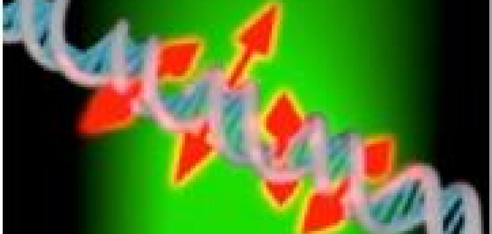
A Stanford University team led by Nobel laureate William E Moerner has developed an enhanced imaging technique for probing the structure of individual DNA strands at the nanoscale. The new method builds on a technique called single-molecule microscopy by adding information about the orientation and movement of fluorescent dyes attached to the DNA strand.
Since DNA is at the root of many disease processes, the new technique could help scientists gain insights into what goes wrong when DNA becomes damaged or when other cellular processes affect gene expression. The work has been published in journal Optica.
Single-molecule microscopy, together with fluorescent dyes that attach to DNA, can be used to better visualise strands of nanometre-thick DNA strands. But until now, it has been difficult to understand how those dyes were oriented and impossible to know if the fluorescent dye was attached to the DNA in a rigid or somewhat loose way.
Moerner from Stanford University is the founder of single-molecule spectroscopy, a method that from 1989 has allowed scientists to visualise single molecules with optical microscopy. Of the 2014 Nobel Laureates for optical microscopy beyond the diffraction limit (Moerner, Hell & Betzig), Moerner and Betzig used single molecules to image a dense array of molecules at different times.
Moerner’s research team demonstrates their new technique by obtaining super-resolution images and orientation measurements for thousands of single fluorescent dye molecules attached to DNA strands.
‘You can think of these new measurements as providing little double-headed arrows that show the orientation of the molecules attached along the DNA strand,’ said Moerner. ‘This orientation information reports on the local structure of the DNA bases because they constrain the molecule. If we didn’t have this orientation information the image would just be a spot.’
Because the technique provides nanoscale information about the DNA itself, it could be useful for monitoring DNA conformation changes or damage to a particular region of the DNA, which would show up as changes in the orientation of dye molecules. It could also be used to monitor interactions between DNA and proteins, which drive many cellular processes.
‘Our new imaging technique examines how each individual dye molecule labelling the DNA is aligned relative to the much larger structure of DNA,’ said Adam S Backer, first author of the paper. ‘We are also measuring how wobbly each of these molecules is, which can tell us whether this molecule is stuck in one particular alignment or whether it flops around over the course of our measurement sequence.’
To acquire single-molecule orientation information, the researchers added an electro-optic modulator to the single-molecule microscope. For each camera frame, this device changes the polarisation of the laser light used to illuminate all the fluorescent dyes.
Since fluorescent dye molecules with orientations most closely aligned with the laser light’s polarisation will appear brightest, measuring the brightness of each molecule in each camera frame allows the researchers to quantify orientation and rotational dynamics on a molecule-by-molecule basis. Molecules that switched between bright and dark in sequential frames were rigidly constrained at a particular orientation while those that appeared bright for sequential frames were not rigidly holding their orientation.
‘If someone has a single-molecule microscope, they can perform our technique pretty easily by adding the electro-optic modulator,' said Backer. ‘We’ve used fairly standard tools in a slightly different way and analysed the data in a new way to gain additional biological and physical insight.’
The researchers tested the enhanced DNA imaging technique by using it to analyse an intercalating dye; a type of fluorescent dye that slides into the areas between DNA bases. In a typical imaging experiment, they acquire up to 300,000 single molecule locations and 30,000 single-molecule orientation measurements in just over 13 minutes. The analysis showed that the individual dye molecules were oriented perpendicular to the DNA strand’s axis and that, while the molecules tended to orient in this perpendicular direction, they also moved around within a constrained cone.
The Stanford team next performed a similar analysis using a different type of fluorescent dye that consists of two parts: one part that attaches to the side of the DNA and a fluorescent part that is connected via a floppy tether. The enhanced DNA imaging technique detected this floppiness, showing that the method could be useful in helping scientists understand, on a molecule by molecule basis, whether different labels attach to DNA in a mobile or fixed way.
In the Optica paper, the researchers demonstrated a spatial resolution of around 25 nanometres and single-molecule orientation measurements with an accuracy of around 5 degrees. They also measured the rotational dynamics, or floppiness, of single-molecules with an accuracy of about 20 degrees.
Related stories
Light-based innovations win Nobel Prizes for Physics and Chemistry
Further information
