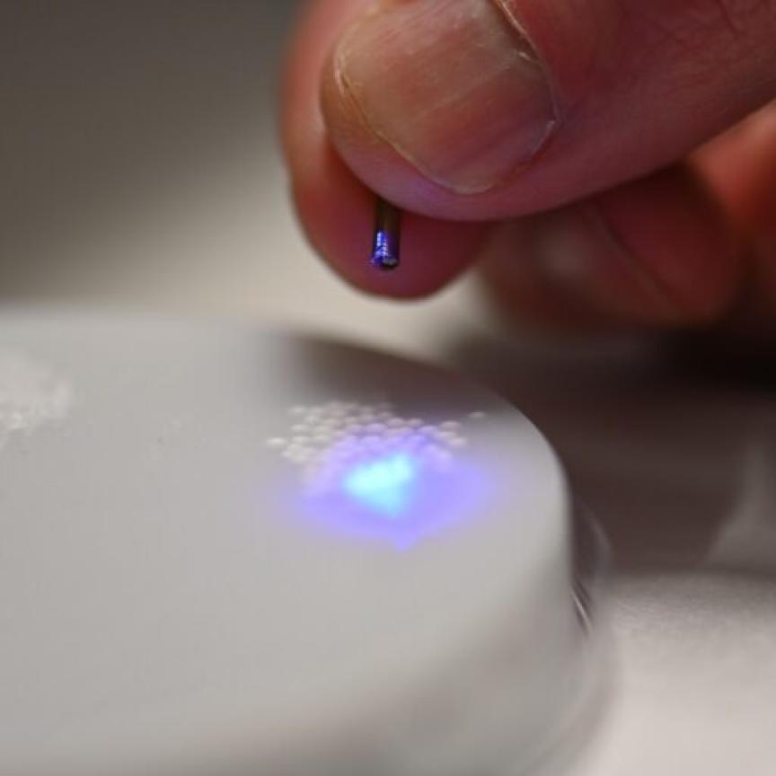
Researchers from the University of Connecticut in the US have used 3D printing to produce an inexpensive, portable and high-resolution microscope that is small and robust enough to use in the field or at the bedside.
The high-resolution 3D images provided by the instrument could potentially be used to detect diabetes, sickle cell disease, malaria and other diseases.
Described by the researchers in Optics Letters, the new microscope – based on digital holographic microscopy – consists entirely of 3D printed parts and commonly found optical components, making it inexpensive and easy to replicate.
According to the researchers, alternative laser sources and image sensors would help further reduce the cost of the system, with a single unit estimated to cost several hundred dollars to reproduce. They added that mass-producing the unit would also substantially reduce its cost.
Escaping the lab
Digital holographic microscopy is typically performed on an optical table in a laboratory, as it usually requires a complex optical setup and a stable environment free of vibrations and temperature fluctuations – which could potentially introduce noise in the measurements. Here a digital camera is used to record a hologram produced from interference between a reference light wave and light coming from the sample. A computer then converts this hologram into a 3D image of the sample.
The new portable system is not only able to perform this technique at a bedside or out in the field, but the 3D images it produces have twice the resolution of those captured using traditional digital holographic microscopy – features as small as 0.775 microns can be resolved. This resolution could be improved even further if a light source with shorter wavelengths were used, according to the researchers.
The researchers were able boost the resolution of digital holographic microscopy beyond what is possible with uniform illumination by combining it with a super-resolution technique known as structured illumination microscopy. This was done through generating a structured light pattern using a clear compact disc.
‘3D printing the microscope allowed us to precisely and permanently align the optical components necessary to provide the resolution improvement while also making the system very compact,’ explained research team leader Bahram Javidi.
Increasing access to healthcare
The new microscope doesn’t require any special staining or labels and could help increase access to low-cost medical diagnostic testing. This would be especially beneficial in developing parts of the world where there is limited access to health care and few high-tech diagnostic facilities.
The current system is ready for practical use, with the researchers planning to use it for biomedical applications such as cell identification and disease diagnosis – continuing their collaboration with their international partners to investigate disease identification in remote areas with limited health care access. They are also working to further enhance the resolution and signal-to-noise ratio of the system without increasing the its cost.
In addition to biomedical applications, the researchers envision the new microscope being used in the research, manufacturing, defence and education sectors.