Researchers have developed a key fundamental technology that can overcome the resolution limitations of existing X-ray microscopes.
The work could help optimise non-invasive nanostructure observation, such as that used in semiconductor quality inspection.
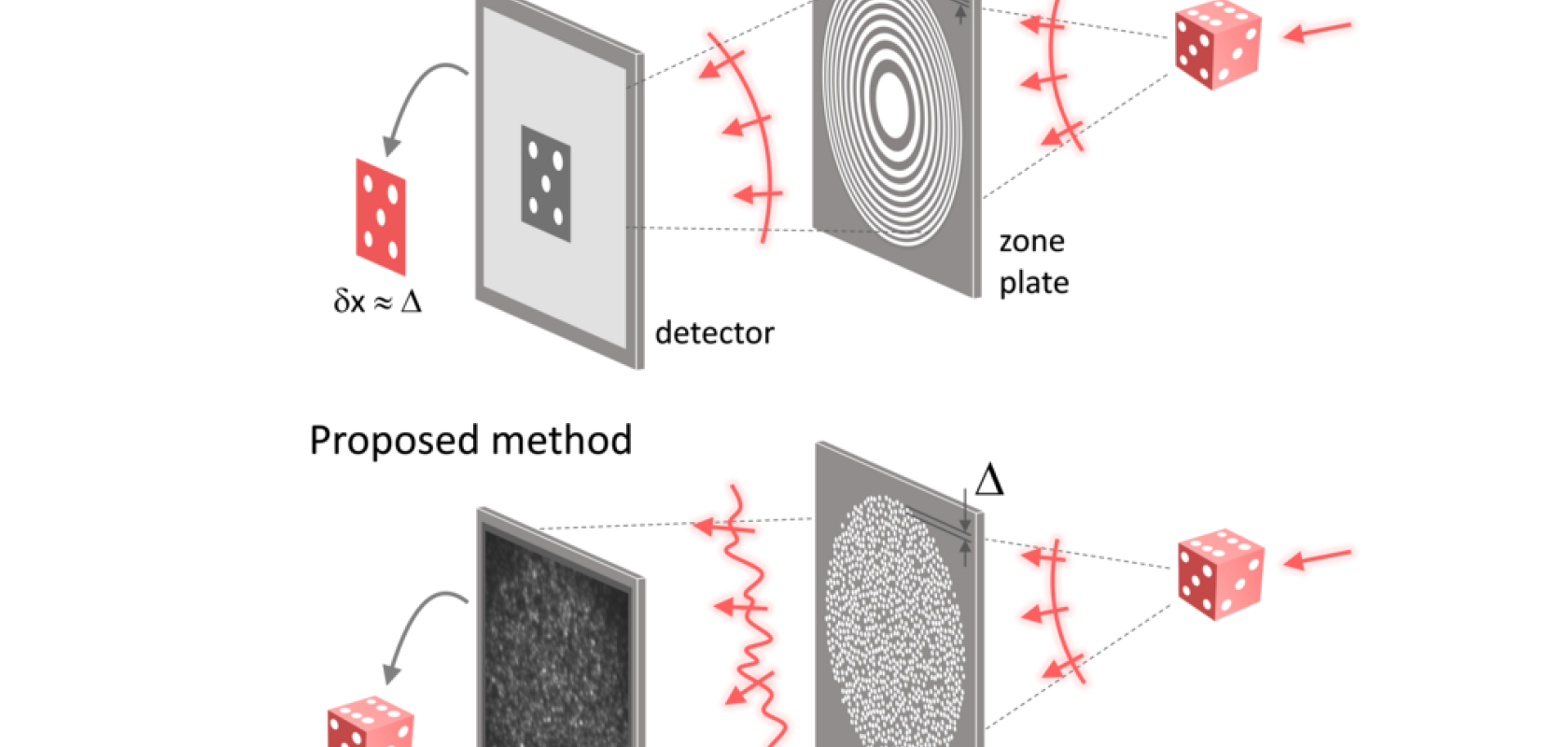
In an X-ray microscope, a circular grating called a concentric zone plate is used instead of a refractive lens. The resolution of an image obtained using the zone plate is determined by the quality of the nanostructure that comprises the plate. However, there are several difficulties in fabricating and maintaining these nanostructures, which set the resolution limit for X-ray microscopy.
In Light Science & Applications, researchers from the Biomedical Optical Laboratory of the Korea Advanced Institute of Science and Technology (KAIST) have proposed a new ‘X-ray lens’ that overcomes these limitations caused by issues in zone plate production.
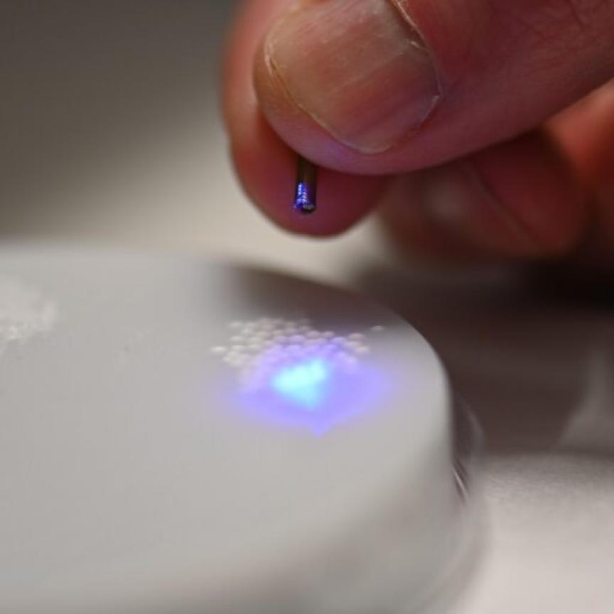
The lens is in the form of a diffuser comprising numerous holes punched in a thin tungsten film, which generates random diffraction patterns by diffracting incident X-rays. The researchers mathematically identified that, paradoxically, the high-resolution information of the sample was fully contained in these random diffraction patterns, and actually succeeded in extracting the information and imaging the internal states of the samples.
The resolution of the image of the constructed sample has no direct correlation with the size of the pattern etched on the random lens used. Based on this idea, the research team succeeded in acquiring images with 14nm resolution by using random lenses made in a circular pattern with a diameter of 300nm.
“In this study, the resolution was limited to 14nm, but if the next-generation X-ray light source and high-performance X-ray detector are used, the resolution would exceed that of the conventional X-ray nano-imaging and approach the resolution of an electron microscope,” said Dr KyeoReh Lee, first author of the Light Science & Applications paper. “Unlike an electron microscope, X-rays can observe the internal structure without damaging the sample, so it will be able to present a new standard for non-invasive nanostructure observation processes such as quality inspections for semiconductors.”
The research was conducted with support from the Research Leader Program and the Sejong Science Fellowship of the National Research Foundation of Korea. More information of the new 'X-ray lens' can be found in the Light: Science and Applications paper.