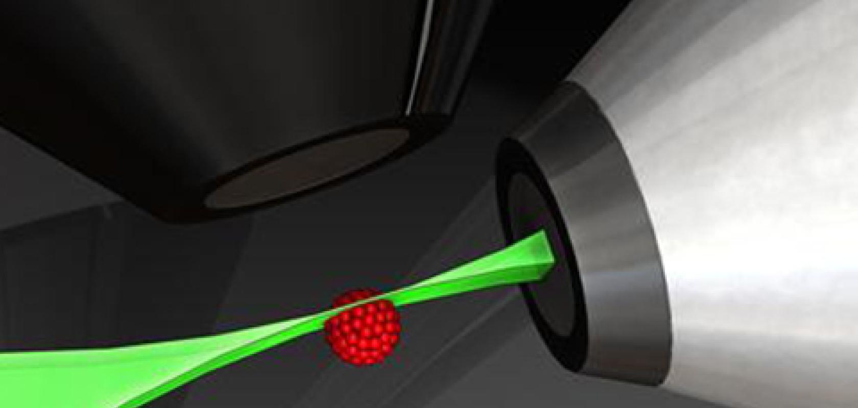
Gemma Church explains how LightTools, from Synopsys, makes tissue modelling more accessible to design next-gen diagnostics.
The interaction of light with biological tissue is notoriously difficult to model. ‘Most users of simulation design packages are accustomed to working with materials that are either very well characterised or easy to measure. Biological tissue is neither,’ Katherine Calabro, senior R&D engineer at Synopsys, explained.
‘It is unfortunately not realistic to import a single material profile with static optical properties and assume it’s “close enough”. Tissue is not a material that has been manufactured to certain specifications in a controlled environment, and as such requires a greater level of knowledge and analysis on the part of the designer,’ she added.
Synopsys is addressing this issue in its LightTools illumination design software. By providing researchers with a tool to accurately model the interaction between light and biological tissue, they hope it will lead to the development of new diagnostic devices and methods.
Brilliant bio-optics
The physics of light interaction with tissue is generally well understood, where scattering and absorption are the two dominant interactions. The effect of scattering can even be further broken down into how often the light is scattered in tissue and also in what direction. This angular distribution of scattering is known as the phase function. Auto-fluorescence is another effect, where light is naturally emitted by some biological tissues.
‘The difficulty, however, is knowing how to measure each of these effects separately,’ Calabro explained. ‘Ultimately, the Holy Grail of a lot of biomedical optics research is knowing how to take an optical signal that is simultaneously affected by all the above interactions, and then decomposing it into separate measurements.’
Traditionally, biological tissue measurements were taken on animal tissues using standard measurement techniques like spectrophotometers. But this work was limited by the fact that the tissue is no longer living. Factors such as the oxygenation of blood, or water draining from the tissue, for example, were not taken into account – but these could have a dramatic effect on the scattering and absorption spectra in living tissue.
‘That is why we have seen such incredible work being done to develop devices and measurement techniques that are able to make measurements in the living body in a repeatable and reliable way,’ Calabro said.
Much progress has been made in this area. Early tissue measurements were only done at a single wavelength. Now, measurements are conducted across the UV to the IR range of the electromagnetic spectrum.
These full absorption spectra can then be further decomposed into the concentrations of chromophores in the tissue: blood volume, blood oxygenation, water content, fat content and so on, allowing optical measurements to be made to identify physiological markers.
Simulation and modelling are also established methods in the field of biomedical optics. A codebase named Monte Carlo for Multi-Layered media (MCML), for example, is a widely shared and used resource for biomedical optics researchers, helping them estimate optical interactions in tissue.
Many of the analysis techniques used to separate complex optical measurements into individual scattering and absorption components use data simulated by these Monte Carlo programs.
But the greatest challenge in the field of biological tissue modelling is variability, according to Calabro. ‘Not only measurement error and variability, but biological variability. Optical measurements can vary widely between different patients, but can also vary widely in the same organ in the same individual. Further, biological tissue is spatially heterogeneous; structures like blood vessels and hair follicles can alter the measurement from one point to another.’
As a result, the values from the same tissue type can vary dramatically, as Calabro explained: ‘Simulations have to make modelling assumptions, not only because it’s impossible to fully characterise the structure of the tissue under the surface a priori, but also because including these small structures dramatically decreases the simulation efficiency. For those reasons, the models have to assume some level of structural homogeneity that is not 100 per cent physically accurate.’
Tissue simulations are also characterised by a high number of scattering events, however a low proportion of rays is actually collected, making such simulations extremely computationally demanding.
‘A single simulation of one scattering value and one absorption value at a single wavelength can take days,’ Calabro explained.
This is where Synopsys’ LightTools can help, providing biomedical optics researchers with a versatile and intuitive tool to design diagnostic methods and devices to measure tissue properties.
‘We have developed a built-in utility that guides the user in building broadband optical property profiles (absorption, scattering, refractive index). It is pre-populated with data and values curated from the research literature. With that as a starting point, the user can modify individual components (such as blood volume or water fraction) as needed,’ Calabro explained.
‘So, even if optically measured data from a particular tissue type is unavailable, users can build a reasonable estimate based on physiologically-based assumptions. These variables can also be used as tolerancing or optimisation variables, with the utility automatically updating the optical properties in the model. These capabilities take tissue modelling beyond simply importing static measured data, recognising the unique challenges that need to be addressed when simulating tissue.’
This provides the end-user with versatility and a comprehensive toolset, where the tools required to design and develop a device with accurate tissue simulation are combined.
Calabro explained: ‘Whereas programs used in academic research settings, such as MCML, primarily use text-based I/O, and are limited to simple geometries like rectangular blocks, LightTools has extensive options for complex geometries and user-friendly analysis tools, with no large text files to parse and analyse separately.’
What’s more, with integrated tools like optimisation and tolerancing, LightTools can properly account for the inherent variability of tissue-optical properties.
This combination of usability and versatility sets LightTools apart from other simulation and modelling tools for those working in the bio-optics field.
In a new technical paper, Synopsys presents the most up-to-date compilation of research and data available in the literature regarding the optical properties of human tissue. It provides the optical, biological and measurement details required when modelling human tissue in LightTools. It also describes how this information has been incorporated into a new Human Tissue Utility. Click here to view the White Paper.
‘While modelling of biological tissue has made tremendous advances in the past few decades, it continues to be a work in progress with many unknowns still to explore. It is our hope that tools such as the tissue material utility in LightTools help make tissue modelling more accessible to those who are not biomedical optics experts. As the field continues to grow, and research data is added to the literature, it is our intention to regularly update and incorporate new data into the utility,’ Calabro concluded.


