‘If you don’t have a cell in the environmental conditions it evolved in, no matter how good your microscope is, how can you be sure that the phenotypes you’re seeing are physiological and real?’ It’s a deeply truth-seeking question that befits the status of its asker, Eric Betzig, as a 2014 Nobel Prize winner in chemistry for developing super-resolved fluorescence microscopy. But now his priority has shifted away from such small scales to getting pictures deep inside living organisms. ‘You need to take the cell and put it back in the system where it evolved,’ continues Betzig. ‘In order to do that and maintain the image quality, you need adaptive optics.’
Betzig, who works at the Janelia Research Campus of the Howard Hughes Medical Institute in Ashburn, Virginia, isn’t the first to use adaptive optics (AO) in microscopy. The approach typically measures distortions in the medium that light travels through, and uses deformable mirrors (DMs) or lenses to shape beams to compensate for it. Adaptive optics has enabled ground-based astronomers to correct for atmospheric effects and, in doing so, see further than ever before. But in seeking to emulate that success, microscopists face difficult decisions in balancing image quality with cost.
‘Adaptive optics is a tool to remove aberrations,’ explains Abdesselem El Hassouni, sales engineer at AO specialist Alpao, in Grenoble, France. ‘In every type of microscopy you have aberrations due to the sample itself. So that means that in every type of microscopy you can use adaptive optics.’ However, microscopy brings significant new challenges. El Hassouni notes that astronomy only considers aberrations during detection. Biological microscopy usually requires illumination to produce fluorescence in a sample, for example by exciting a fluorophore. It’s therefore important to shape light’s illumination wavefront and get precise excitation, and then also consider aberrations during detection.
El Hassouni explains that microscopy systems can use DMs to correct for aberrations in open or closed loops. Open loop control reshapes DMs into iterative or pre-determined geometries, while closed loop calculates geometries based on feedback from wavefront sensors. Closed loop is precise, but users can’t always get enough photons to the wavefront sensor to enable it, El Hassouni says, plus wavefront sensors add cost and complexity to the optical path. Open loop systems therefore use their initial imaging scan to determine which preset geometry the DM should adopt. Alpao has developed its own open loop-based plug-and-play AO system that does this, which can be combined with any microscope.
Betzig says that it was obvious to him that AO would be valuable to neuroscientists when he started at Janelia in 2006, ‘because of the aberrations that they face in imaging in the brain’. Together with his wife Na Ji, who has pioneered AO approaches, their work initially focused on two-photon fluorescence microscopy[1]. This technique is popular because it can excite fluorophores using two infrared photons arriving simultaneously, scanning samples with light beams rather than completely flooding them with light. In part because infrared photons scatter less in tissue, this approach offers deeper penetration than other techniques, up to around a millimetre in depth.
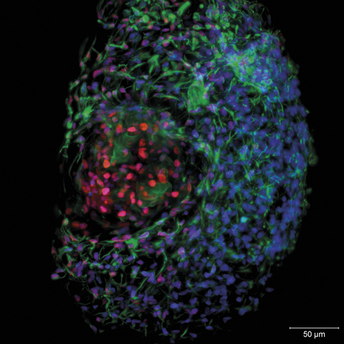
Zeiss' classical light sheet microscopy provides better understanding of hepatocyte spheroids, a model system for drug-induced liver injury, liver function and disease. Credit: TNG Weiquan John, Ng Huck Hui, Genome Institute of Singapore
‘Eventually, in 2014, we published a paper about a two-photon guide-star-based approach for doing confocal and two-photon microscopy,’ Betzig recalls[2]. This uses two-photon fluorescence throughout a sample, creating single spots as guide stars, and measures resulting aberrated wavefronts with Shack-Hartmann wavefront sensors.
Characterising aberrations mathematically to correct them using AO involves Zernike polynomial functions. Simpler corrections can use low-order Zernike modes, whereas more difficult ones require many more, high-order modes. Some existing AO microscopy techniques deal only with the low-order Zernike modes to avoid wavefront sensors. ‘But when you’re trying to image 200µm deep in a zebrafish, the amount and complexity of aberration is far higher than what most AO methods have been trying to correct,’ Betzig says. ‘The wavefront sensing approach that we use handles up to 55 or more Zernikes.’
Seeing clear benefits
Such method development has some commercial two-photon microscope vendors showing increasing interest. Thorlabs Imaging Systems in Sterling, Virginia, for example, works closely with Ji and Betzig. ‘We definitely are looking at adaptive optics and have built and demonstrated a multiphoton system using adaptive optics,’ reveals Sam Rubin, Thorlabs Imaging Systems’ general manager. ‘But we don’t yet, as of November 2018, offer it as a commercial option in our system.’ He expects such an AO tool would benefit in vivo neuroscience most, for example by enabling imaging deeper into tissue. ‘The deeper you go, the more you’re starting to see the effect of scattering in the tissue,’ Rubin says. ‘Na Ji’s work has shown that adaptive optics can significantly improve that.’
Rubin notes that Thomas Bifano, at Boston University, has shown that combining multiphoton microscopy with AO can also make it easier to study the brain[3]. ‘You can image directly through the skull, and that simplifies preparation work and improves success rates of experiments,’ Rubin says. ‘We are doing some work with academic collaborators on that.’
Thorlabs does, however, already supply AO kits, adds John Taranto, advanced imaging research engineer, which can include DMs developed by Boston Micromachines Corporation (BMC), co-founded by Bifano. Such kits can also include Shack-Hartmann sensors, ranging from lower cost and lower frame rate products to higher frame rate CMOS sensors that enable faster closed-loop control. Thorlabs also produces its own piezoelectric-based DM, with a 10mm diameter active area and 32 actuator segments, suitable for handling lower Zernike orders. BMC’s silicon MEMS-based electrostatic actuator DM, with a 12x12 actuator grid array in a 4.4mm2 area, corrects for higher spatial orders as well.
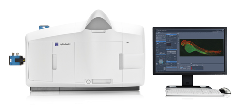
Zeiss' Lightsheet Z.1 system can already adopt some adaptive optical correction measures. Credit: Zeiss
But another important factor is the DM’s stroke, the deformation distance they can achieve. ‘The Boston Micromachines MEMS structures’ stroke is about 3.5µm, which is short compared to our piezo-based system,’ Taranto says. ‘Depending on how many Zernike modes you’re deploying, it could have a min-max stroke distance of 13µm.’ Thorlabs is also currently developing a product optically conjugating both DMs, so that one corrects low-order aberrations and the other mirror corrects higher-order aberrations, Taranto adds.
Alpao’s DM69 deformable mirror offers very large strokes of around 60µm, says El Hassouni, which enables microscopes to image much deeper inside tissues. Betzig’s team uses that capability to address many Zernike modes in its closed-loop systems. But El Hassouni adds that Alpao’s 69 ‘silicon spring’ actuators are also highly stable, made of the same material as the 10.5mm diameter mirror itself. That is valuable in open-loop systems, where the mirror must reliably form pre-set shapes.
Academic groups can get the microscopes HHMI develops from Betzig, and Alpao has an agreement to supply DMs to these new microscope owners at a preferential price. However, El Hassouni admits that its DMs are a ‘little bit overspecified’ for many applications, making them a luxury for those only correcting low-order aberrations. Therefore, although Alpao is an industry leader in AO DMs, it still sells less than 100 per year.
Alpao is therefore developing a mid-range DM that will correct about 10 Zernike modes, and be more affordable. These low-order DMs will be specially built to have very low wavefront error, like the DM69, for those modes. It is also integrating the DM’s driver electronics, which are currently a separate 2kg unit, with the mirror itself in a single module weighing 200-300g. ‘We are looking for partners, who are saying “in three to five years, I will need 100 pieces”,’ El Hassouni says.
Thorlabs offers two different types of deformable mirrors in its adaptive optics kits, which can handle different numbers of Zernike modes. Credit: Thorlabs
The DM69 mirror has enabled Betzig to integrate AO into another technique his team developed at the same time as its guide-star method: lattice light sheet microscopy (LLSM)[4]. Light sheet microscopy, in general, images biological systems fast but gently, providing 3D information over wide fields of view with high spatial and temporal resolution. It illuminates samples with thin ‘light sheet’ laser beams, covering only in the sample portion being imaged. Systems then collect the resulting fluorescence in a perpendicular direction from the illumination axis. LLSM uses one light beam divided into seven parts, exposing samples to minimal radiation, preventing damage and accelerating the scanning process.
Biological response
To integrate with LLSM, AO ‘needs to be fast and photon efficient’, Betzig explains, so that the sample’s fluorescence is still evident after correcting for aberrations. ‘The initial microscope is a Frankenstein graft of three microscopes: an adaptive optics for extinction, an adaptive optics for detection and a lattice light sheet in the middle,’ he says. ‘It’s expensive and technically complex, because we require an ultrafast laser to create a two-photon guide star and it requires a wavefront sensor, and also an expensive camera and other optics. In my own opinion that cost and complexity is worth it though because it’s fast, non-invasive, and allows you to correct for really messy aberrations.’ In their April 2018 Science paper, Betzig’s team used the approach to show cancer and immune cell movement and nerve circuits wiring up in ‘unprecedented 3D detail’[5].
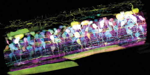
Using adaptive optics with lattice light sheet microscopy, Eric Betzig’s team can see inside the spinal cord of a zebrafish embryo as new neurons light up in different colours and track nerve circuit development. Credit: T. Liu et al./Science 2018
Betzig has licensed the basic non-AO LLSM approach to microscopy giant Zeiss, based in Jena, Germany, which has in turn sublicensed the technology to Intelligent Imaging Innovations (3i), in Denver, Colorado. 3i is the only commercial LLSM system, according to Tiemo Anhut, staff scientist for laser scanning microscopy methods at Zeiss Research Microscopy Solutions. ‘At the moment LLSM is certainly a rather advanced method in the field of live cell imaging,’ says Anhut.
‘We do believe, however, it will become a commodity in the cell biology field as soon as turnkey systems become available.’
Meanwhile Zeiss offers its Lightsheet Z.1 instrument commercially for the more classical form of light sheet microscopy. ‘It is the ideal system to observe living specimens and follow their development over long periods of time and capture highly dynamic processes,’ Anhut says. ‘Moreover, images of samples such as mouse brains can be acquired at high resolution and speed.’ This contrasts with LLSM, which typically focuses on cells and small organisms.
Zeiss has not implemented any adaptive optic components like DMs in the detection arm in its Lightsheet Z.1 instrument yet, Anhut says. ‘In the end, one has to judge if simpler corrections can be sufficient,’ he explains. ‘There are various methods to perform adaptive optical correction. It is possible, for instance, to use Zeiss objectives with motorised correction collars on our classical microscope stands, with which one might influence the wavefront, depending on the imaging depth within the sample. Certain adaptations to the sample under study are possible in the Lightsheet Z.1 too, such as the optimal positioning of the light sheet relative to the observation objective.’
While Anhut calls integrating AO into LLSM ‘a tremendous improvement’, he has reservations about its cost and complexity. ‘One will have to carefully balance the benefits, compared to the efforts one has to take at which site the AO is introduced in the system,’ he says. Pioneering labs like Betzig’s always push technology limits, he adds. ‘It is the task of companies like us to make such complex technologies available for non-experts, and hence implement them in a stable and user-friendly way,’ Anhut says. ‘But certainly AO will have an impact on imaging deeper into the specimen, and its demand will grow with the development of more and more sophisticated imaging systems like the LLSM.’
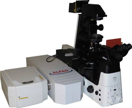
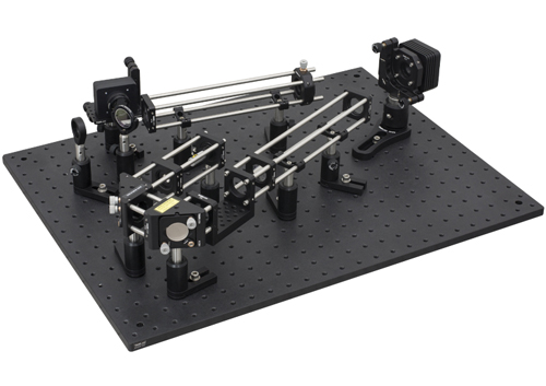
Alpao’s adaptive optics system sits between the microscope on the right and the optical setup on the left enabling approaches like multiphoton or lattice light sheet microscopy. Credit: Alpao
Meanwhile, Betzig’s team is taking advantage of needing an instrument with AO and ultrafast lasers for the guide star, along with many other components used in microscopy. They are trying to make a ‘Swiss army knife’ instrument that can incorporate several modes of operation. But of the functions it will offer, Betzig has no doubt which is most significant.
‘The lattice is probably the most important technology I’ve developed in my career,’ he stresses. ‘It’s easily the one that has gotten the best response from the biologists, even more so than the PALM technique that got me the Nobel, because it offers super high speed, it’s really good, near isotropic resolution and extremely low photodamage. But to really take advantage of that in a multicellular context, you need the adaptive optics.’
Andy Extance is a freelance science writer based in Exeter, UK.
References
[1] N Ji et al Curr Opin Neurobiol 2008 DOI: 10.1016/j.conb.2009.03.009
[2] K Wang et al Nat Methods 2014 DOI: 10.1038/nmeth.2925
[3] D Sinefeld et al Opt. Express 2015 DOI: 10.1364/OE.23.031472
[4] B-C Chen et al Science 2014 DOI: 10.1126/science.1257998
[5] T-L Liu et al Science 2018 DOI: 10.1126/science.aaq1392
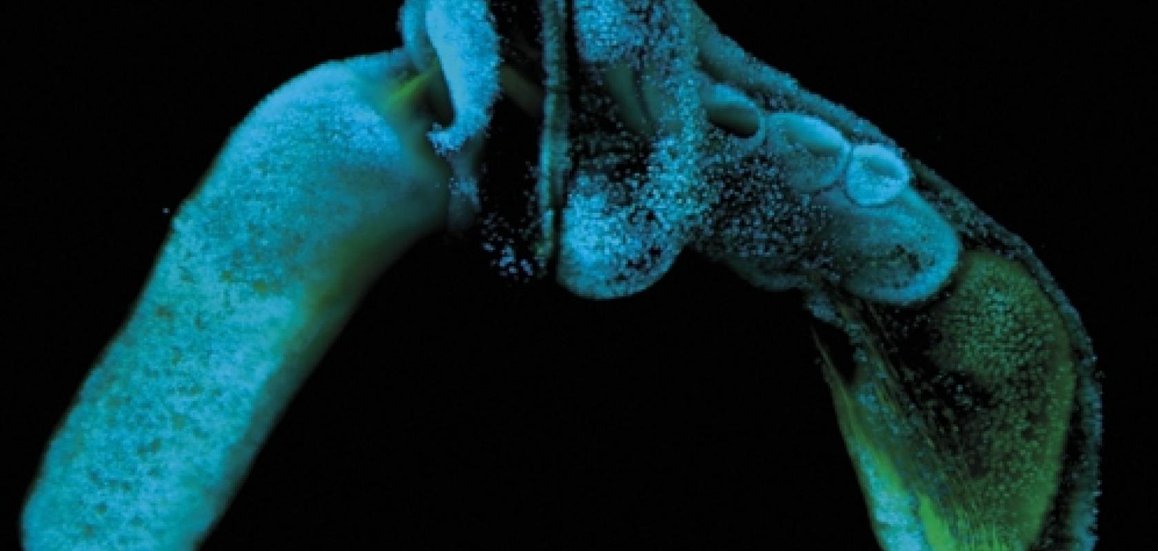
Top image: Fluorescently labelled acorn worm (Saccoglossus kowalevskii) imaged with a Zeiss Z1 light sheet microscope. Part of research by Dr Jessica Gray, working in Marc Kirschner’s lab in the systems biology department at Harvard Medical School. Credit: Jessica Gray, Harvard Medical School / Zeiss
