It’s almost one hundred years since Raman spectroscopy was first discovered, but it is only with recent technological developments and falling prices that we are finally beginning to see its potential realised in a host of different fields. Applications of Raman spectroscopy are as diverse as tracking corrosion, identifying diseases, and helping in the production of pharmaceuticals.
At 2022's Laser World of Photonics, Professor Dr Jürgen Popp of the Leibniz Institute of Photonic Technology told attendees about work to combine different imaging techniques – such as hyperspectral imaging, coherent anti-Stokes Raman scattering, second harmonic generation microscopy, or two photon fluorescence microscopy – into a multi-modal imaging approach that takes the best parts of each one.
Speaking to Electro Optics, Popp noted that some of the most exciting developments in Raman spectroscopy are where they are beginning to be translated into medical applications. He gave an example of being able to use Raman spectroscopy for cancer diagnostics, to give the type, stage and grade of cancer.
‘We can also use Raman spectroscopy for rapid identification of infection-causing pathogens, for determining the antimicrobial resistance against certain antibiotics,’ he said. ‘We can determine the minimal inhibition concentration of these antibiotics, or if the bacterium has already developed a resistance against the antibiotics.
‘We can also determine the host response,’ he continued. ‘So let’s assume a patient comes into the doctor’s office with typical symptoms of an infection. Raman spectroscopy can be used to quickly determine if this really is an infection or not, or if it is something like a sterile inflammation. And, if it is an infection, then we can even use Raman to see whether it is a viral infection, a fungal infection, or a bacterial infection, just by looking at the Raman chemical fingerprint of white blood cells. We have now reached the stage where we can translate these findings step by step into the hospital, into the practice, and also into the market.’
Professor Popp is involved in Leibniz Centre for Photonics in Infection Research (LPI), which aims to accelerate the development and commercialisation of light-based diagnostic methods and novel therapeutic approaches for treating infectious diseases. For instance, one of the centre’s research projects is to develop methods and technologies that can detect viral outbreaks earlier and support their containment, to help society be better prepared for future epidemics.
In terms of Raman spectroscopy, Professor Popp, working with colleagues Shuxia Guo and Dr Thomas Bocklitz through the LPI centre, published a guide on Raman spectral analysis in Nature Protocols. The researchers want to contribute to standardised Raman spectral analysis to make it easier to apply in medical or biological settings. The guide covers steps from experimental design and data preparation to data modelling and statistical analysis, while pointing out potential pitfalls and how to avoid them.
Elsewhere, Professor Popp said he had recently been working on using Raman spectroscopy for early detection of neurodegenerative disease by examining the eye. He said that Raman spectroscopy has been shown to be able to differentiate between mice exhibiting Alzheimer’s disease and those that do not by imaging the retina. This approach has now been transferred within a European project called Moon into a medical device to record Raman spectra of the retina in-vivo. Currently the Moon device is being tested on Alzheimer’s patients in the medical university of Vienna.
Advantages of Raman
Raman spectroscopy is a way of gathering chemical information about a substance by how light scatters at different wavelengths. Shine a laser on a target, such as a bacteria or even virus particles, collect the inelastic scattered light, and that will give a molecular fingerprint of the sample. In comparison to other spectroscopy methods it has the advantages that it is relatively simple to use, provides a lot of information, and, importantly, does so non-invasively.
Professor Popp explained: ‘With some mathematical algorithms you can easily interpret this molecular fingerprint and either identify those cells or learn something about what is going on in the cell.
‘The big advantage is it's a label-free technique,’ he added. ‘You get the information directly on the native sample, and you can do the experiments in such a way that you can look at living cells without disturbing them, which makes it completely different from many other techniques like fluorescence [spectroscopy], for example, where you need external labels to achieve molecular selectivity.’
Also speaking about the advantages of Raman spectroscopy, Dr Dieter Bingemann, senior application scientist at Wasatch Photonics – which makes Raman spectrometers – commented: ‘The output from Raman spectroscopy is very specific. It has a lot of information; it can tell apart every compound in the world from the spectrum. Anything that's semi-transparent the laser can go through and information comes right back. So you can have instant chemical analysis inside the container in a second by shooting a laser at it.
‘Other spectroscopy methods often need a path – an absorption path, an input and output – which limits how you can make measurements,’ he continued. ‘In Raman, you can just immerse your probe in the sample, or interrogate and collect information from the same side. It has all the practical advantages needed to be widely applicable across disciplines.’
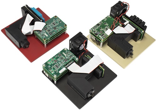
Raman spectrometer modules from Wasatch Photonics, which can be integrated into many application-specific systems including medical devices. Credit: Wasatch Photonics
These practical advantages have increased significantly with the huge reduction in both size and cost. As the portability and costs have fallen, the range of applications have risen, and as Michael Matthews, VP of spectroscopy at Wasatch Photonics, explained, it’s particularly useful for dealing with the messy world of biology. ‘Historically, Raman has been used in areas like security and defence applications where people are trying to mask or cut a drug or explosive into various mixtures to hide it,’ he said. ‘Now we see that same power to pull out target molecules from an unknown matrix being applied to biochemical and biomedical applications.
‘Biology is very messy,’ Matthews added. ‘It changes from sample to sample, it changes from moment to moment, and a lot of spectroscopy techniques struggle with that. But Raman is particularly good at getting meaningful results from very complex and messy samples, which makes it ideal for use in point-of-care settings, from rapid screening to pathogen detection.’
Bingemann also highlighted the biotechnological applications of Raman spectroscopy. In particular it is already being seen in large fermenters used to cultivate biopharmaceuticals such as antibiotics, therapeutic proteins, enzymes and insulin. ‘In fermenters it’s essential to control the glucose levels delivered to the bacteria, to keep the glucose low but constant, and that's one thing you can do well with in-situ Raman spectroscopy,’ he said. ‘In the past, once you closed the chamber and turned on the feeding motor, you had no further control over the output. Now you can measure the glucose level instantly and adjust on-the-go to increase both the output and quality substantially while reducing process time and waste.’
Realising Raman’s potential
The potential of Raman technology is seemingly limitless, although for widespread adoption devices need to get smaller and lower cost. There have already been rapid improvements in that direction, as Matthews explained, but there can still be some societal barriers. ‘There aren't too many diagnostics that are utilising lasers [on] humans, particularly the direct in-vivo measurements,’ Matthews said. ‘Our customers that are sending Raman-based medical devices through clinical trials are constantly asked to address this laser interaction, laser safety. That's something that may slow down the eventual adoption, but from a hardware and a science point of view it's a very clear pathway to this becoming a reality.’
Matthews highlighted two other developments driving the adoption of Raman technology: firstly, AI and new ways of pulling information out of data streams, which, he said, is ‘going to really unlock the potential of Raman’; and secondly, the availability of processing power.
The widespread application of Raman spectroscopy is also likely to have a part to play in driving its adoption as it is likely to contribute to a virtuous circle of falling prices and rising adoption. Matthews commented: ‘The biggest opportunity is not a single application but actually the aggregate of all the potential. If you look at the type of new applications under development, any single application alone could be bigger in market potential than the total Raman market is today. Raman is poised on a precipice where any one of these could take off, driving the volume up and bringing the cost of the equipment down, which will allow even more applications. I foresee some breakthrough applications in the near future – particularly in medical diagnostics – that will make Raman a game-changing technique over the next decade.’
Almost one hundred years after Raman spectroscopy was discovered and its potential is only just beginning to be realised. What is clear, however, is how much potential there is.
--
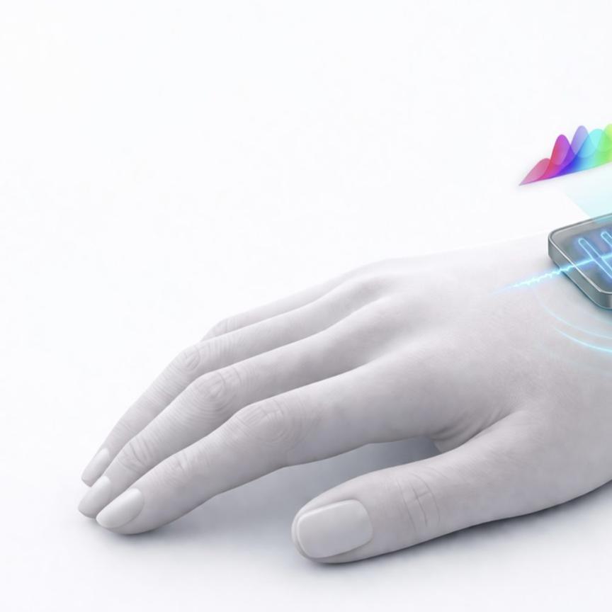
Gold Raman wearable sensor designed for skin biomarker analysis
Researchers at the University of Tokyo have created a wearable sensor, spun from gold, that can measure different biomarkers on the skin using Raman spectroscopy. The work was published in Advanced Optical Materials.
Shining a laser light from a Raman spectrometer onto the sensor can give information about the chemicals present on the skin at that point in time. The sensor can be tuned to be extremely sensitive, and is robust enough for practical use.
The main component of the sensor is a fine gold mesh. Gold is unreactive so it doesn’t affect biomarkers in sweat, for instance. In addition, the mesh is so fine that it provides a large surface area for a biomarker to bind to, the chemical composition of which can be detected by a Raman spectrometer.
‘Currently, our sensors need to be finely tuned to detect specific substances, and we wish to push both the sensitivity and specificity even further in future,’ said assistant Professor Tinghui Xiao. ‘With this, we think applications like glucose monitoring, ideal for sufferers of diabetes, or even virus detection, might be possible.’
‘There is also potential for the sensor to work with other methods of chemical analysis besides Raman spectroscopy, such as electrochemical analysis, but all these ideas require a lot more investigation,’ said Professor Keisuke Goda at the University of Tokyo. ‘In any case, I hope this research can lead to a new generation of low-cost biosensors that can revolutionise health monitoring and reduce the financial burden of healthcare.’
Sponsored: Compact laser modules aid Raman spectroscopy instrument OEMs
Raman spectroscopy is a non-destructive optoelectronic analytical technique used to determine the chemical composition of materials. It utilises the inelastic (Raman) scattering of light from chemical bonds within a sample to identify molecules by their unique Raman spectrum. This spectrum consists of a number of peaks showing the wavelength and intensity of the Raman scattered light, with each peak caused by intra- or inter-molecular vibrations. The Raman spectrum of each molecule is unique and can be used like a fingerprint as identification.
Raman scattering theory
When light interacts with a molecule, the incident photon excites the electron cloud of the molecule, which raises its energy into a virtual energy state. This virtual energy state is unstable and so almost immediately afterwards a photon is re-emitted as scattered light. Almost all of the scattered light has exactly the same wavelength as the incident photon and the energy of the molecule remains unchanged. This elastic scattering is named Rayleigh scattering after the 19th-Century British physicist, Lord Rayleigh (John William Strutt).
A tiny fraction of the scattered light exhibits a different wavelength to that of the incident photon. In these incidents, the photon has either imparted energy to the molecule and the resultant scattered photon has a longer wavelength (lower energy), or the molecule relaxes to a lower vibrational level and the scattered photon has a shorter wavelength (higher energy). This inelastic process is known as Raman Scattering after the Indian physicist, C. V. Raman.
Applications
Raman spectroscopy systems can provide a quick and convenient method of sample identification since they require minimal sample preparation. It is even possible to perform Raman spectroscopy through containers, which enables applications from aviation security to pharmaceutical analysis. Another advantage of Raman spectroscopy is that water molecules do not produce Raman scattering, and so chemicals or biological samples contained within water can easily be detected.
Raman spectroscopy systems require a very stable, high-power, narrow linewidth light source to create sufficient Raman scattered photons for detection. The quantity of photons that are Raman (inelastic) scattered are tiny compared to Rayleigh (elastic) scattered, at approximately 1 in 10 million! It is therefore important to separate these two forms of scattered light to enable the detection of the relatively weak Raman signal.
Lasers
Laser Components supplies high power, narrow linewidth and very stable lasers, such as the NovaTru Chroma series modules, as light sources for Raman spectroscopy to OEM instrument manufacturers globally. The NovaTru Chroma laser modules use a high-performance Volume Bragg Grating to ‘lock’ a conventional laser diode to a very narrow linewidth with high wavelength stability (± 0.5nm), which is virtually temperature independent (typically 0.01nm/°C). A monitor photodiode and thermistor are used to regulate the light output with automatic power control feedback. These modules are relatively compact and cost- effective when compared to traditional gas lasers.
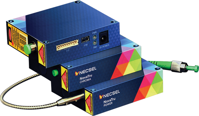
The NovaTru Chroma laser modules use a high-performance Volume Bragg Grating to ‘lock’ a conventional laser diode to a very narrow linewidth with high wavelength stability
The most popular wavelengths used for the excitation light source in Raman spectroscopy systems are 780, 785 and 830nm. Higher Raman scattering cross-sections are achieved with shorter wavelength lasers, however, there is a trade-off, since background fluorescence also increases with shorter wavelengths. The company offers suitable modules for all common Raman spectroscopy wavelengths.
Optics
The Rayleigh scattered light corresponding to the laser line is typically filtered out using a notch filter, edge pass filters or bandpass filters, leaving only the wavelength shifted Raman scattered light, which is collected and dispersed onto the sensor. Laser Components manufactures and supplies the complete set of optics used in Raman spectrophotometers, from lenses and beamsplitters to optical filters and diffraction gratings, customised for individual system requirements. Also offered are a range of optical subassemblies, such as digital monochromators, housing the entrance slit, mirrors and tuneable grating in a single unit, ideally suited for general spectroscopy use, as well as OEM system integration.
Further information www.lasercomponents.com/uk/product/wavelength-stabilized-laser-source/