Researchers in the United States have developed a UV light-enabled catheter for fixing congenital heart defects non-invasively. In the near future, the device, which has been described in Science Translational Medicine, could enable doctors to fix holes in the heart without needing open surgery.
The UV catheter was designed as part of a joint effort between Boston Children’s Hospital, the Wyss Institute for Biologically Inspired Engineering at Harvard University, the Harvard John A Paulson School of Engineering and Applied Sciences (SEAS), and the Karp Lab at Brigham and Women’s Hospital.
A hole in the septum of the heart – the inner wall dividing the left and right sides of the organ – is a common type of congenital heart defect. The faulty septum effects blood flow through the heart allowing oxygen-rich blood to mix with deoxygenated blood.
The catheter, however, could represent a radical change in the way these kinds of cardiac defects are repaired. ‘In addition to avoiding open heart surgery, this method avoids suturing into the heart tissue, because we’re just gluing something to it,’ said Pedro del Nido, MD chief of cardiac surgery at Boston Children’s Hospital and contributing author on the study.
Catheterisations are preferable to open heart surgery because they don’t require stopping the heart, putting the patient on bypass, or cutting into the heart.
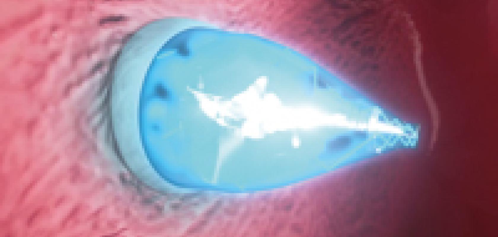
The catheter uses UV light to place an adhesive patch inside the beating heart. First, the UV catheter is directed non-invasively through a vein to reach the defect. There, the catheter is pulled back to reveal a device that uses small expanding balloons to seal off the hole in the tissue. The balloons deploy biodegradable adhesive patches, and the UV light from the catheter reflects from the balloon’s shiny interior and activates the patch’s adhesive coating. As the glue cures, pressure from the positioning balloons on either side of the patch help secure it in place. Then, the device and catheter are gently extracted, leaving the patch in place.
Not only does the device negate the need for open surgery, but the adhesive patches support tissue regrowth before they biodegrade naturally. This is more beneficial than traditional surgery involving stitches that remain in the body, as these can move out of place or fail to cover the hole as the body grows.
‘This really is a completely new platform for closing wounds or holes anywhere in the body,’ said Dr Conor Walsh, contributing author on the study, assistant Professor of Mechanical and Biomedical Engineering at SEAS, and founder of the Harvard Biodesign Lab at SEAS. ‘The device is a minimally invasive way to deliver a patch and then activate it using UV light, all within a matter of five minutes and in an atraumatic way that doesn’t require a separate incision.’
The device has also been designed to be customisable, meaning that the rate at which the patch biodegrades can be slowed or accelerated depending on how quickly the surrounding tissue grows over it. According to the authors of the study, further research will reveal the appropriate lengths of time depending on different circumstances.
Dr Jeff Karp, a bioengineer at Brigham and Women’s Hospital and a co-founder of medical device company Gecko Biomedical, developed the glue product in his lab at Brigham and Women’s Hospital. Gecko Biomedical will be testing the glue product in humans later this year.
‘Our collaboration across hospitals and institutions to find new and minimally invasive applications for this glue in clinical settings is a great multi-disciplinary example,’ Karp said. ‘We are translating our discoveries in the lab into solutions for patients.’
‘The way the glue works in the face of blood is revolutionary. We don’t have to stop the heart,’ added del Nido. ‘This will enable a wide range of cardiac procedures in the future.’
