Photonics technologies are allowing scientists to move ever closer to understanding how the brain works, and what can go wrong during destructive diseases such as Parkinson’s and Alzheimer’s. One exciting area of research, optogenetics, is allowing scientists to selectively control neural activity with pulses of light.
At the same time, advanced optical microscopy techniques such as two-photon microscopy are allowing neurobiologists to image this neuronal activity more precisely and at deeper points within the brain. The inventors of the technique, Winfried Denk and Arthur Konnerth from Germany, and Karel Svoboda and David Tank from the USA, were presented with a €1 million European neuroscience award, the 2015 Brain Prize, on 7 May at a ceremony in Copenhagen, Denmark.
‘The combination of CW lasers for neuron activation and inhibition with two-photon imaging techniques has allowed neurologists to activate and simultaneously image neural pathways, allowing them to map neural networks and to better understand their individual functions,’ explained Barney Mitchell, vice president of sales and marketing at Laser Quantum. ‘This is pushing forward our knowledge of neurological diseases, and will ultimately lead to better treatment and possibly cures for diseases such as Parkinson’s.’
Interest in these techniques has increased following the launch of the BRAIN initiative (Brain Research through Advancing Innovative Neurotechnologies) in the United States in 2013, which aims to ‘revolutionise our understanding of the human brain’.
In September 2014, a further $300 million of funding was announced for the project, $30 million of which was committed to the US photonics industry for the development of new optics and photonics technologies. Although too early to notice any effects in the private and commercial sector, the programme is leading to a surge of activity in brain research from academic institutions and scientists, according to Stephan Briggs, biomedical engineer at Edmund Optics. ‘In universities and research labs we’re seeing a tonne of people kick off these newer projects that tie to things like optogenetics… or brain tissue imaging.’
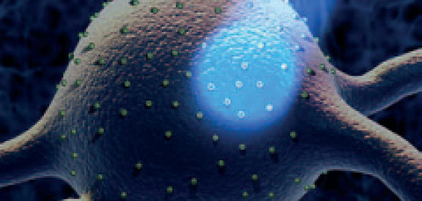
In order to ‘control’ neural activity, neurons are first treated with light-sensitive proteins known as rhodopsins. Channel rhodopsin (ChR2), for instance, responds to blue light, so a 473nm continuous wave laser can activate the neurons treated with this protein. ‘If you have this neuron in the front of your brain, in the prefrontal cortex, you’re able to shine blue light onto it, which essentially drives activation,’ explained Briggs. ‘So, if we know someone is deficient in a certain electrolyte or mineral, we can use blue light to activate a neuron which then signals the brain to pump more of this chemical into the body.’
Alternatively, cellular expression can be repressed through the use of proteins known as halorhodopsins, which respond to light in the green part of the electromagnetic spectrum.
In order to deliver light into the brain and activate neurons, different types of optics can be used depending on the size of the area being targeted. For smaller areas, scientists can use probe style objectives, which require micro optics on the order of hundreds of microns up to one or two millimetres in size in order for them to be inserted into the brain. For larger areas, the laser can be coupled to a fibre optic. ‘A fibre optic would have a very large dispersive area, so you could cover more neurons and activate more channels. If it was a smaller area, then an objective would be use to focus light more accurately,’ said Briggs.
Although optogenetics is still in the laboratory stage and has not yet been applied to humans, continually advancing technologies as well as increased funding and public interest will only help push it to the next stage. ‘Getting this technology into industry is the first and foremost thing that we need to do − right now it’s all in pre-clinical trials. I think the BRAIN initiative will be one of the things to help drive this further,’ commented Briggs.
Two-photon microscopy
During optogenetics studies, as well as being treated with light-sensitive proteins, neurons are also loaded with a fluorescent dye that binds to calcium, so that the neuronal activity can be monitored by two-photon imaging of the fluorescence-tagged calcium.
Two-photon fluorescence microscopy, the inventors of which won the Brain Prize in May, has become a transformative tool in brain research, allowing scientists to examine three-dimensional images of biological specimens in real time. The function of individual nerve cells as they communicate with each other in networks can now be studied, which is a huge step forward in the understanding of the physical processes of the human brain, as well as the mechanisms behind neurological disease.
The technique uses an advanced form of fluorescence microscopy, whereby cell components are labelled with molecules that fluoresce under ultraviolet light, which are then observed using a microscope. The process involves exciting a fluorophore from the electronic ground state to an excited state by a single photon. However, the short-wavelength, high-energy UV-light used for this more conventional method spreads through the tissue, making it difficult to focus upon a specific cell or cell part
Two-photon microscopy uses the same excitation process, but through the simultaneous absorption of two less energetic photons, typically in the infrared spectral range under sufficiently intense laser illumination. Infrared light can penetrate deeper into the tissue, meaning that scientists can observe real changes inside a living, active brain, even down to hundreds of micrometres below the brain surface. And, unlike conventional fluorescence microscopy, the infrared light does not exhaust the fluorescent molecules.
Advances in ultrafast laser sources have played a significant role in enabling these multiphoton techniques that allow for imaging at greater penetration depths and with lower phototoxicity. ‘Femtosecond lasers are fast changing from huge, complicated instruments requiring constant maintenance, suitable only for optical physicists, to compact, sealed systems with user-friendly software, well suited to biologists as a simple tool rather than a project,’ remarked Mitchell from Laser Quantum.
Because excitation of the fluorescent dye depends on the simultaneous absorption of two photons, the intensity of the infrared light needs to be extremely high. Therefore, ultrafast laser pulses − typically with pulse durations of less than 200fs − are needed in order to deliver this high-intensity light in suitably short pulses to avoid overheating the sample.
One of the most popular fluorescent dyes used in two-photon microscopy is GFP – green fluorescent protein − and the most commonly used absorption peak for GFP is at approximately 470nm. A suitable laser for two-photon microscopy needs to be roughly twice the wavelength of the absorption spectrum, so that two photons at the infrared wavelength (in this example, at around 940nm) can combine to give the equivalent energy of a single visible (470nm) photon.
Depending on the individual application, there is often a compromise to be made for the repetition rate of the laser. Typically, a few milliwatts or tens of milliwatts are required at the sample, Mitchell pointed out. Higher power will give a brighter image by increasing the peak power of the femtosecond pulses, but this also increases photo-bleaching and cell damage, resulting in greatly decreased imaging times. Longer pulses reduce the peak power, but this decreases the probability of two photon excitation, so duller images are achieved and performance becomes similar to that of a lower power, short pulsed laser. ‘This is where a 1GHz rep rate offers a clear advantage over the traditional 80MHz rep rate, as multiplying the repetition rate by 12 gives the option of 12 times the fluorescence for the same peak power, or a similar level of fluorescence with lower peak powers. This reduces cell damage and photo-bleaching, giving a far longer imaging time,’ explained Mitchell.
The beam of a femtosecond laser is sent through free-space optics, and is sometimes shared between multiple microscopes − as powers are generally 1W or greater at the laser, and at most tens of milliwatts are used at the sample − and then directed through the microscope and down onto the sample.
In two-photon microscopy it is necessary to take into consideration the dispersion of ultrashort pulses in the microscope optics. While passing though microscope optics, the low-frequency part of the femtosecond laser pulse travels faster than the high frequency. As a result, the short pulse becomes longer, with the low-frequency part arriving first to the specimen. This effect is referred to as ‘chirp’.
‘When the original pulse length is in the order of 100-150fs, there is little dispersion, and so the pulse remains useably short at the sample,’ explained Mitchell. ‘Where shorter pulses are used, sub-50fs or so, the bandwidth of the pulse is broader, and so the dispersion due to the optics, particularly the objective lens of the microscope, leads to a long pulse at the sample. Dispersion compensation is therefore required for short pulsed lasers before the pulse is sent through the objective lens, to counter this effect. There are a range of commercially available pre-chirpers for this purpose.’
But while lasers are advancing, objective lenses and coatings are also being improved to achieve the best light transmission in the microscope.
‘Sometimes lasers can have properties which would cause a normal system to fail,’ remarked Briggs from Edmund Optics. ‘One of those could be extremely high power… or they could have a very fast pulse repetition… people are pushing the envelope with femtosecond pulses. The spot sizes can be also incredibly small − so with a high power, high pulse rate and small spot size, you get a very large energy density in that spot size. So, our lenses need to be able to withstand those densities.’
Anti-reflective coatings are essential for any lens to prevent diffraction and energy loss, but companies are expanding the spectral correction over a broader range of wavelengths for use in more advanced microscopes. ‘We’ve really paid attention to having that spectral correction from 400nm to 1,500nm,’ said Briggs. ‘This is absolutely critical for two-photon or multi-photon microscopy; you’re typically exciting with some type of near infrared or infrared wavelength, which could be anywhere from 800nm to 1µm, or the Nd:Yag band and its harmonics – 1,064nm, 532nm, 355nm, 266nm.’
Objective lenses are also starting to incorporate fluorides, particularly calcium fluoride, for their high spectral transmissive properties from UV wavelengths all the way to infrared wavelengths, Briggs added.
 Jessica Rowbury is a technical writer for Electro Optics, Imaging & Machine Vision Europe, and Laser Systems Europe.
You can contact her on jessica.rowbury@europascience.com or on +44 (0) 1223 275 476.
Find us on Twitter at @ElectroOptics, @IMVEurope, @LaserSystemsMag and @JessRowbury.
