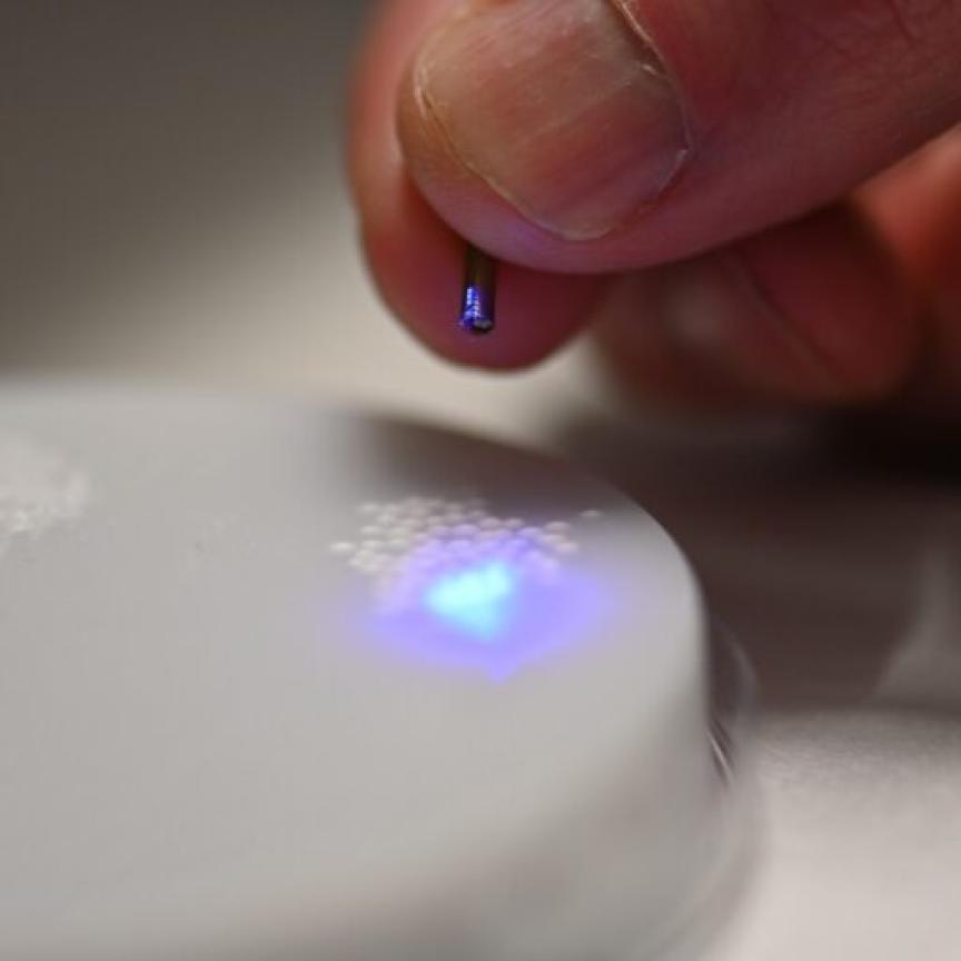
A new microscope has been developed at Queen Mary University of London’s Blizard Institute that opens up new opportunities in biological imaging. The spinning disk super resolution imaging (SDSI) microscope is able to produce pictures with a resolution of 80nm and allow artefacts such as protein complexes in the nucleus to be viewed, which currently can’t be achieved using standard PALM and STORM super-resolution techniques.
The team that developed the microscope was led by Dr Ann Wheeler, head of imaging at the Blizard Institute, and Professor Martin Knight of Queen Mary’s School of Engineering and Materials Science.
The new SDSI microscope was developed by combining the single focus plane technology of a spinning disk system with the super resolution techniques of Photoactivation Light-Microscopy (PALM) and Stochastic Optical Reconstruction Microscopy (STORM). It allows imaging in live cells, which other super resolution microscopy techniques would find difficult.
Dr Wheeler, Prof Knight and their team used piezo-driven, capacitive sensor feedback nano-positioning technology from Physik Instrumente (PI) to provide long-term stability for the SDSI microscope.
Currently PALM and STORM super-resolution techniques use TIRF to collect fluorescent images over an extended time period and then use complex algorithms to process the data. The high laser power required for TIRF illumination can cause photo-bleaching in live cells, which limits the technique’s range of use. Also, the penetration depth of TIRF is limited to 100nm; therefore preventing access to the cell nucleus.
The microscope at the Blizard Institute uses a spinning disk to acquire the image and then processes the data using super resolution algorithms. Acquiring the images using the spinning disc allows both a lower laser power to be used and penetration depths to reach the level of the nucleus. The technique produces pictures with a resolution of 80nm and can be used to view protein complexes in the nucleus.
Each set of data taken from a single image plane is acquired over several minutes. During data collection the sample must remain stable relative to the focal plane. Any drift in the microscope due to thermal effects must be compensated for. Despite leaving the microscope to stabilise over several hours and keeping it in a temperature controlled environment, the scientists at Blizard were seeing drift in the focal plane.
The P-725 PIFOC piezo driven nanopositioning objective drive was used in the system, which incorporates PI’s own capacitive position sensor to provide position control and stability to the nanometre scale over a wide temperature range.
The capacitive sensor measures the air gap between two plates of the same material placed only a few tens of microns apart. The design minimises the effects of temperature change ensuring nanometre precision is maintained over a wide operating range. Once fitted to the microscope the PIFOC was successfully used to compensate for drift during the measurement cycle. Implementation of the PIFOC has provided the scientists at Blizard with the stability required in the SDSI to generate an extensive range of data.
The data has shown the viability of SDSI as an enhancement to existing techniques through the ability to collect super resolution images with good signal-to-noise ratio in any selected axial plane within a cell.