A team of international researchers have developed a spectroscopic point-of-care device that could enable consistent and cost-effective screening for thyroid nodules. Funded under Horizon 2020, the technique could lead to faster diagnosis and prevent unnecessary surgeries.
The team are to present the project at the OSA Biophotonics Congress: Optics in the Life Sciences meeting, which will take place in Florida between 3 and 6 April.
Early diagnosis in thyroid cancer can improve a patient’s likelihood of recovery, but current screening methods use instruments with poor sensitivity and can yield inaccurate results. Consequently, doctors often have to rely on incomplete information to make diagnostic decisions and recommend treatments, and this can lead to patients receiving unnecessary surgeries or experiencing a reduced quality of life.
Currently, standard thyroid screening methods involve an initial ultrasound with sub-optimal sensitivity and resolution. If the ultrasound detects an abnormal nodule, clinicians perform a fine needle aspiration biopsy (FNAB), to test for malignancy. But, FNAB results often do not lead to a diagnosis or are false positives. These inaccuracies can subject patients to unnecessary surgeries.
‘The problem is in the poor specificity of the current approaches which leads to a significant number of unnecessary biopsies and surgeries,’ said Turgut Durduran, the project coordinator and professor at the Institute of Photonic Sciences (ICFO), Barcelona, Spain. ‘Unfortunately, current imaging or screening modalities are not able to distinguish malignant nodules from benign nodules with a good specificity.’
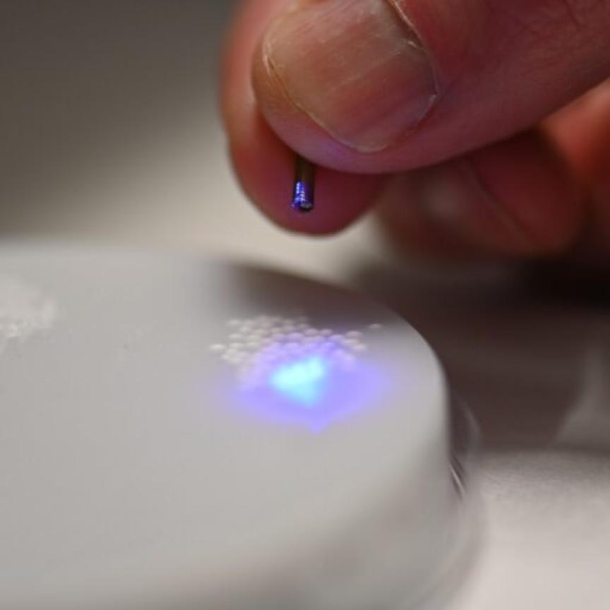
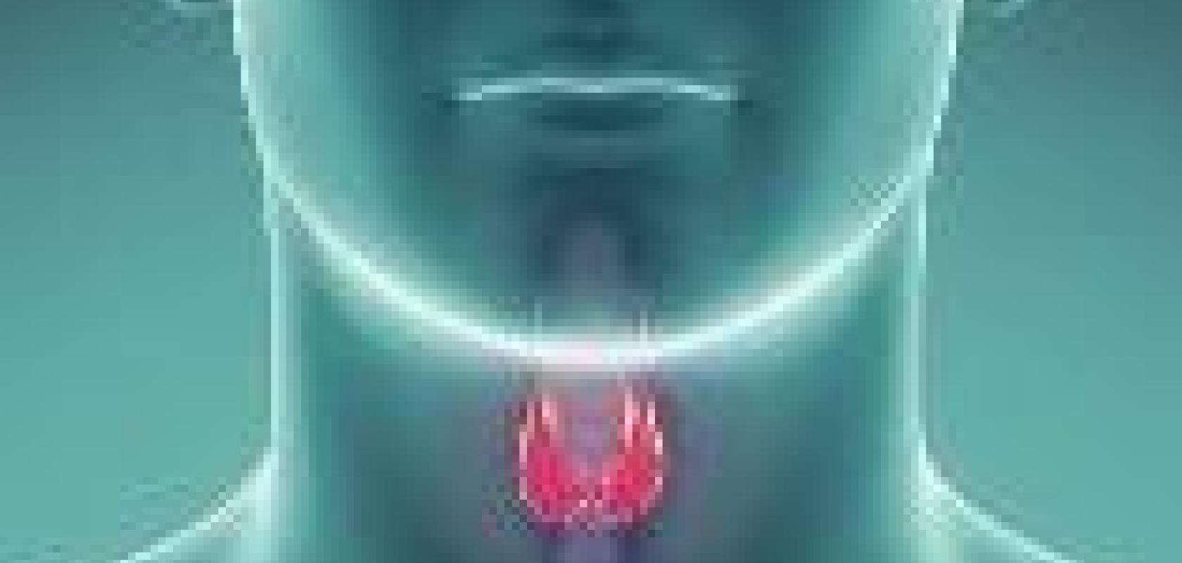
The project, called ‘Laser and Ultrasound Co-analyser for thyroid nodules (LUCA), aims to develop a technology that improves data acquisition for medical professionals by probing chemical constitution, water concentration, structure and haemodynamics – like blood flow and oxygenation – of tissue simultaneously. This device builds on the current ultrasound standard with a hybrid optics/ultrasound probe.
The device uses near-infrared time-resolve spectroscopy (TRS) and diffuse correlation spectroscopy (DCS) to collect all the tissue data, each independently a commercial-level technology already. The DCS laser subsystem features a fibre-coupled laser diode at 785nm and custom-developed driving and cooling electronics. The custom design cuts the device cost by 10 to 15 times that of a standard DCS laser system.
The optical module also collects data on chromophore concentrations, like water and lipids, through TRS. The TRS subsystem, which features photomultipliers and time-correlated single photon counting, also cuts the cost to about five times lower than commercially available equivalents.
According to the team, the high prevalence of thyroid nodules, at up to 76 per cent of the population, means that even modest strategy improvements for characterising lesions could have a major positive impact. And in fact, they have already seen how this optical innovation could impact patients’ lives if it were in the clinic.
‘In a pilot study, the mere fact that the ultrasound screening was carried out next to our measurements identified a malignant nodule in a healthy, young volunteer, and we have seen that many nodules that went all the way to a surgery turned out to be benign,’ said Durduran.