A group of Italian researchers has developed a modification to optical mammography instrumentation that increases the sensitivity of the medical imaging technique by up to a thousand-fold. The researchers reported their research at the OSA Biophotonics Congress ‘Biomedical Optics’, which took place 3-6 April in Hollywood, Florida, USA.
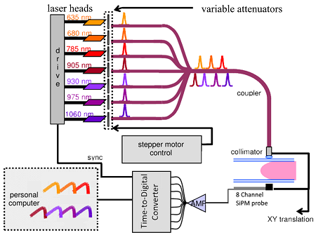
Schematic Diagram for OM Instrument: Seven pulsed lasers sequentially illuminate the compressed breast; transmitted light is detected by the 8-channel SiPM probe and the TDC acquires the signal. (Image: Ferocino et al.)
Optical mammography (OM) uses harmless red or infrared light to image tissues and can be used in conjunction with x-rays to reduce the amount of ionising radiation patients must be subjected to when diagnosing and monitoring cases that require repeated imaging. The technique has attracted increasing interest in breast cancer diagnosis thanks to its two wavelengths being highly sensitive to the composition of tissue. However, so far only poor spatial resolution has been achieved when using it.
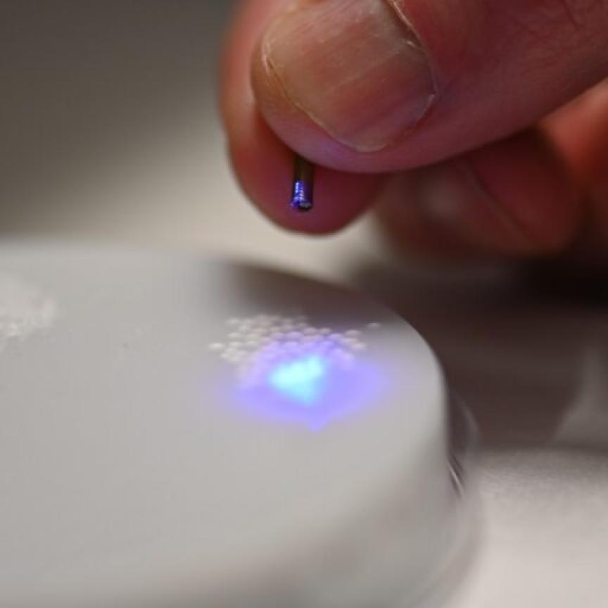
The Milan-based researchers’ new instrument design replaces two photomultiplier tubes of existing OM instruments with an eight-channel probe featuring silicon photomultipliers and a multichannel time-to-digital converter. Not only do these changes eliminate a time-wasting pre-scan step that was originally required to avoid damage to the photomultiplier tubes, but they also increase the sensitivity of OM by up to a thousand-fold while making the equipment cheaper and more robust.
While the poor spatial resolution of OM does still remain a challenge when used as a stand-alone technique, it has shown promise when combined with other imaging methods, for instance it could be used to measure the blood volume, oxygenation, lipid, water and collagen content of suspicious tissue areas previously identified by x-ray imaging. A tumour for example would be characterised by a high volume of blood due to the increased vascularisation that occurs as it grows.
While x-ray mammography is still the recommended method for routine breast cancer screenings – many of the estimated 252,710 breast cancer diagnoses last year were made using it – the technique suffers from low sensitivity (only achieving 50-75 per cent), uses potentially harmful ionising radiation, and is limited by a patient’s age, weight, body mass index and breast tissue itself.
Considerable pressure of the breast tissue is also required when using x-ray mammography, whereas OM requires only gentle pressure of the tissue when performing imaging. Three-dimensional OM detectors are also being developed that will eventually require no pressure on the tissue at all, according to the researchers, which would instead surround the tissue with rings of light sources and detectors when imaging.
OM also shows promise for use in pre-surgical chemotherapy, according to Milan-based researcher Edoardo Ferocino: ‘This technique is able to provide information on the outcome of chemotherapy just weeks after beginning treatment, or possibly even sooner.’ The Italian research group is therefore planning clinical studies to further explore the use of OM to monitor and predict the outcome of chemotherapy.
The researchers are also working with a larger consortium on a project known as SOLUS, ‘Smart Optical and Ultrasound Diagnostics of Breast Cancer’. This project is funded by the European Union through Horizon 2020 and aims to combine optical imaging methods with ultrasound to improve specificity in the diagnosis of breast cancer.
Related articles
Micro-optical X-ray sensor to enable high-precision medical imaging
Photonics takeaways from the Medica trade fair
Photonics-guided robots to transform operating rooms, experts say