Gemma Church explains how a new Raman hyperspectral spectrograph unlocks a fast chemical analysis technique
Many spectroscopy techniques now exist for a range of applications. Vibrational spectroscopy is one such analytical tool, allowing fast and non-destructive tests without the need for extensive sample preparations.
There are a number of vibrational spectroscopy techniques to choose from, including Raman spectroscopy, which is a non-invasive chemical analysis technique.
Here, a molecule scatters incident light from a laser source and a tiny proportion of the scattered light – the Raman spectrum – reveals detailed information about a sample's chemical structure and molecular interactions. The Raman spectrum features a number of peaks where each peak represents a specific molecular bond vibration.
Often, a Raman spectrum is referred to as a chemical fingerprint of a particular molecule or material. As such, Raman spectroscopy is a highly desirable technique across applications where non-destructive, chemical, microscopic imaging and analysis are required.
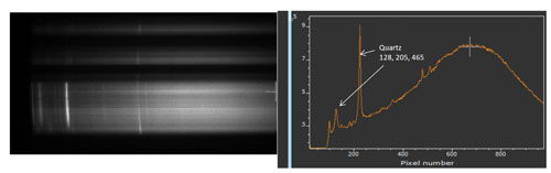
Single frame showing Quartz Raman spectra
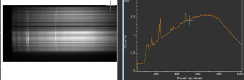
Single frame showing multiple Raman spectra of albite, dolomite and quartz
Speedy spectroscopy
Raman microscopy is one of the most popular analysis techniques that relies on Raman spectra, where you place a sample on the microscope stage and then scan across a range of points to complete the analysis.
But you have to measure one point at a time and while this Raman spectroscopy works well for certain applications, it becomes infeasible if you need a high throughput where, for example, you need to scan multiple samples quickly.
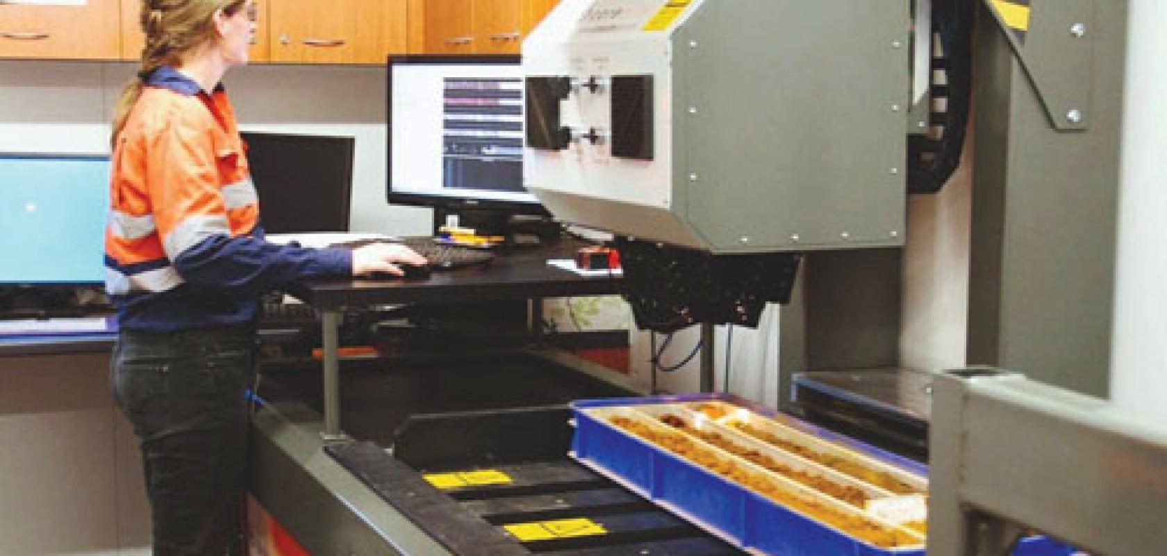
This is where Raman hyperspectral systems can help. Mike Sullivan, founder and president of SensIR, said: 'With a typical Raman microscope, you may be able to measure an array of 75x75 points in a couple of minutes. With our Raman hyperspectral system, you can measure thousands of points within 20 milliseconds. The amount of data this system is capable of generating is humongous.'
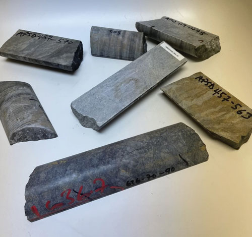
A selection of mineral samples
Over the last 35 years, SensIR has developed a number of hyperspectral imaging solutions across the visible-near infrared (VNIR), near-infrared (NIR) and short-wavelength infrared (SWIR) range. It is a manufacturer for UK supplier Photonic Solutions, which specialises in providing cutting-edge optoelectronic products for research and industry.
SensIR's hyperspectral solutions are now found across a range of applications. This includes chemical imaging for medical applications, where hyperspectral imaging is an emerging diagnostic technique in disease diagnosis and image-guided surgery.
Hyperspectral imaging is also used when processing food, helping sorters to identify and remove any non-compliant food, defects or foreign materials that a traditional camera may miss. Such systems can identify the fat content in meats, characterise powders and grains, and identify spoiled foodstuffs and pathogens.
Airborne hyperspectral imaging is another popular option in the agricultural industry, where these cameras are attached to drones and used for crop identification, soil health monitoring and other analysis applications.
Mineral mapping is another popular area, where hyperspectral units in the 400 to 2,500nm wavelength range are used to analyse drill core, rock chips and other geological samples for the mining, oil and gas industries.
SensIR is currently developing a series of mid-wavelength infrared (MWIR) hyperspectral cameras for the 2,500 to 5,000nm range. Its Raman hyperspectral instruments are also available as a complementary solution to its traditional long-wavelength infrared (LWIR) solutions. There is a slight difference between LWIR and Raman spectroscopy – LWIR spectra show the absorbance of specific molecular bond vibrations. But, with Raman, the spectra are of emitted vibrations.
Sullivan explained: 'Our Raman products came about because we were trying to develop mid- and long-wave infrared hyperspectral solutions, which is very challenging. Raman is complementary to LWIR. So, if the rock samples have signatures in the LWIR, they're going to have them for Raman hyperspectral too.'
Raman is a good alternative to LWIR spectroscopy. For example, water can interfere with LWIR-based measurements, but Raman remains unaffected. 'So, it's a more sample-friendly option, compared to LWIR,' Sullivan explained.
'The Raman hyperspectral system is also not plagued by many background issues seen with other systems. An LWIR spectrometer has to manage large thermal backgrounds with cooled housings, low spectral resolution and expensive IR cameras, [for example].'
But Raman spectrometers often do not provide the performance required for real-world scenarios. These limitations include low optical throughput, a limited area of interrogation due to the focused laser spot size, and large size and weight requirements. Plus, many such systems use focused laser excitation, which can cause eye safety concerns, restricting the use of Raman spectrometers for most real-world applications.
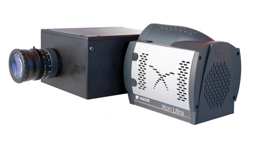
SensIR's 532nm Raman hyperspectral solution addresses these issues. It is an F/.95 spectrograph design and uses an EMCCD camera to capture every photon.
'Getting enough signal-to-noise in tens of milliseconds was a challenge during the Raman hyperspectral system's development,' Sullivan explained.
'Also, we are using a high-power green laser, spreading it out on the line we want to measure. Everything has to be enclosed around the system.'
F1 Raman hyperspectral camera
One of the stand-out advantages of the SensIR system is its high throughput, providing real-time scanning capabilities over a 2-inch span with integration times as low as 20 milliseconds.
The SensIR Raman spectrograph also has a spectral resolution of 0.5 cm across a 25-micron slit. The enclosed system also operates at a standoff distance of up to 250mm between the system and the sample. It is also only 4kg in weight, with dimensions of 75 x 150 x 150mm, making it suitable for a wide range of applications.
In the company's latest White Paper, SensIR reveals how its Raman spectrograph was used to identify core samples containing quartz, albite, pyrite, rutile, white mica, calcite and dolomite minerals, which were moving at a rate of 25mm per second.
The system's design is based on a traditional push-broom sensor concept, but using hyperspectral imaging over 256 spatial channels with a spatial resolution of 0.25mm on a strip of 64mm width. A spectral resolution of better than 6mm was achieved across the 100 to 2,000nm range.
'Our Raman hyperspectral system provides users with everything they need to chemically analyse their samples with speed and peace of mind, thanks to SensIR\'92s 35 years of experience developing next-generation hyperspectral imaging solutions,' Sullivan concluded.
For more information on this topic please click here to download a White Paper.