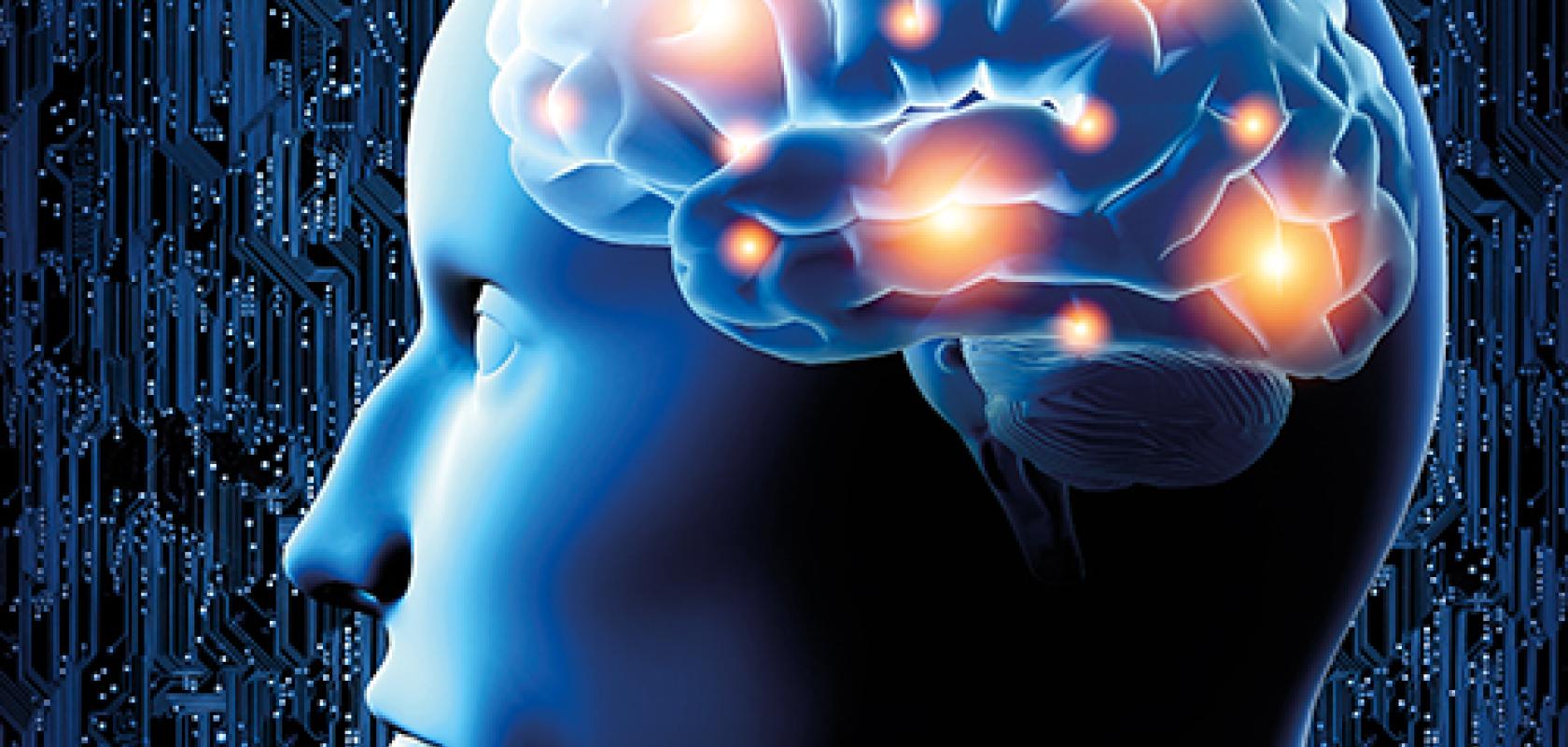
Neurophotonics, described as the application of light-based tools and technologies to observe the finest details of the human nervous system and brain, is advancing brain research and the development of therapies for neuro-diseases and psychiatric disorders.
Valentin Nägerl, professor of neuroscience and bio-imaging at the University of Bordeaux, noted that the development of new kinds of bio-sensors (and actuators), advanced microscopy techniques and image analysis tools among other elements is helping to push the broad and dynamic field of neuroscience forwards.
However, further improvements across every area are needed to advance the scope and usefulness of neurophotonics methods. Research groups across the globe are working on such developments, integrating elements of optics, biology and physics to craft ever better technologies to penetrate ever deeper into living tissues such as brain.
While substantial funding programmes like the BRAIN initiative put the US in a strong position for neuroscience research, other regions are also excelling in the development of neurophotonics instruments and techniques.
With a history of manufacturing optical systems and pioneers like Leica and Zeiss, Germany is at the forefront of the commercialisation of super-resolution microscopy techniques, which have made it possible to study the complex and dynamic inner life of cells at the level of the individual protein building blocks. Meanwhile, in France scientists have been focusing on biological applications and discoveries enabled by these new techniques, Nägerl pointed out.
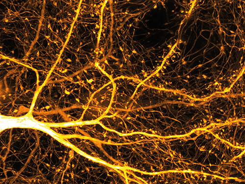
Of the many emerging developments in neuroscience, Nägerl considers the new generation of super-resolution light microscopes to be a breakthrough. By improving the ability to look deep inside biological tissue, these microscopes for instance allow taking extremely sharp and detailed images of neurons and their activity in real time as the animal subject goes about its everyday tasks.
This discovery brought professor Stefan Hell, director at the Max Planck Institute for Biophysical Chemistry in Göttingen, and his colleagues a Nobel Prize in 2014.
By breaking Abbe’s limit, the diffraction barrier in optical microscopy, Hell was able to increase the power of resolution tenfold, and he also showed there was room to obtain even better resolutions.
‘These advances in imaging make it possible to study, in real time, molecules and processes within the brain that have so far been out of reach for non-invasive optical methods,’ commented Nägerl. ‘We can now see things at the nanoscale, way beyond the old textbook limit set by the diffraction of light. And we can do so not by just scratching at the surface but really looking deep into it.’
The higher spatial resolution allows scientists to see molecules, for example, as they move dynamically in a natural context in real time.
‘The more we can see and understand, the more hope there is that we will be able to find cures and therapies,’ said Nägerl. ‘There is always a thirst to see more.’
Neural highway
A traffic management structure can be used to demonstrate how neurons in the brain communicate. City traffic systems can flow very smoothly, but they can also malfunction. Traffic lights can fail in one place. A car accident can lead to a jam in another. In the same way, neurons can fail to flow as they should or make proper connections. Malfunctions in neuron communications and processes inside our brains can lead to degenerative diseases such as Alzheimers and Parkinsons. But now that advances in neurophotonics allow scientists to see the mechanisms behind such neuronal degeneration, they are much better placed to correct processes and find cures.
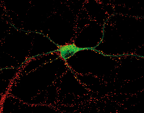
Optogenetics is one neurophotonics technique that allows scientists to use light to observe, or even control, the activity of neuronal circuits in order to better understand how the brain works.
‘Akin to the different traffic lights and signals that allow to control or direct the flow of traffic circulation on a road, neurophotonics [specifically optogenetics] can also be used to up or down-regulate the activity of a specific subset of neurons, using genetically engineered light-sensitive proteins,’ explained Mario Méthot, coordinator at the Neurophotonics Center of the Canadian Neurophotonics Platform.
‘As for now, [optogenetics] is applied in basic science research… but scientists have great hopes that knowledge acquired using model systems could eventually be applied in humans to treat various diseases affecting the brain,’ said Méthot.
Marie Carlen, associate professor of Neuroscience at the Karolinska Institute at the University in Solna, Sweden, is one research who is studying living animals using optogenetics techniques.
Laser and fibre optics are used to ‘insert’ varying intensities of light to be directly into specific areas in the brains of experimental animals, which causes specific neurons to be turned on and off.
The corresponding changes observed in the animals’ behaviour give clues as to which cells and regions of the brain are involved in different emotional and physical responses, such as those associated with hunger, fear, anxiety, learning, motivation and more.
This element of control happens because of opsins, the molecules in neurons that can be activated by light. Some types of opsins fire off positively charged ions in response to light, making it possible to activate or switch on neurons. Other opsins, by contrast, send out negatively charged ions when in contact with light, and inhibit or turn off neurons. Genetic engineering has allowed opsins to be tailor-made to fulfill specific functions and better meet the needs of researchers seeking clues as to which neuronal functions correlate with behaviours. It is already the case that light can be used to manipulate opsins with a high degree of precision allowing for neurons in specific locations to be turned on and off for specific lengths of times.
The lasers and optics needed for optogenetic experiments are specialised. Marie Carlen works with Swedish photonics firm Cobolt to develop the laser devices that best enable her team to advance their studies of the cognitive functions of animals. Calibrating laser tools so that they deliver sufficient light to the area of an animal brain to switch specific neurons on and off without damaging the tissue is a complex task, involving the correlation and adjustment of many different factors such as wavelength, power output and stability. In addition, the laser devices have to be flexible enough to move as the animals move. Cobolt offers a variety of user-friendly lasers to meet the various needs of researchers such as Marie Carlen.
In other research, Benjamin Judkewitz, professor in Bioimaging and Neurophotonics at the NeuroCure Cluster in Berlin, is developing a technique to track and image neurons as they interact with each other and communicate inside their natural networks, potentially allowing scientists to model brain activity in real time, as subjects perceive multiple real world phenomena simultaneously and respond. This technique would help scientists, for example, track the emergence and growth of a tumor in a particular location and develop therapies. Immunologists, on the other hand, may be able to study the process by which lymph nodes become swollen – and not just cells in a petri dish.
The sheer complexity of the brain and nervous system, however, makes the task of modelling them a long term project. The brain alone has about 86 billion neurons. Tracking them as they interact with each other to generate complex and ever changing patterns of movement as they respond to their environment is a staggering challenge.
To go back to the metaphor of the traffic system, Judkewitz and his team seek to monitor and model the trajectory not just of one vehicle moving inside a city, but the movement of all vehicles, airplanes and pedestrians in all the cities and transport systems across world at same time and all in real time multiplied by a factor of a billions and all this, on a molecular scale.
The limitations of optical microscopy make the challenge even more difficult. When light is scattered and defracted, it results in the destruction of the information that light photons have been carrying. Judkewitz is developing an optical time reversal technique to counteract the effect of light scattering and to harvest more information from photons. His approach involves using a microscope to send photons back along the path they have travelled, back in space and time, to reproduce what exactly it was that a photon encountered and saw when it was inside tissue.
With so many breakthroughs on so many fronts, there is every reason to be excited about the possibilities of the huge and ever expanding field of neurophotonics. Not only can it help answer fundamental questions of science such as those concerning the nature of consciousness, commented Professor Nägerl, but also be used to solve real-world problems, for example, cures for degenerative diseases such as Alzheimers.
