In May 2020, Spanish police discovered an international gang smuggling cocaine into Europe from Colombia by impregnating it into cardboard used in boxes carrying pineapples and limes. Once received in Europe, chemists could extract the cocaine from the cardboard.
This continues a trend towards drugs being distributed by embedding them in paper, explained Sulaf Assi, an analytical chemist at Liverpool John Moores University. Assi has been using Raman spectroscopy to detect whether drugs are embedded into a piece of paper or card. ‘I could detect some of the drugs which had been impregnated, but there was a challenge with the sensitivity,’ she said.
Assi is one of many scientists around the world seeking to use Raman and infrared spectroscopy to solve forensic puzzles. She and other scientists have successfully developed methods to find evidence to help solve crimes, where the insights they can provide are remarkable. In many cases they can detect illegal drugs, determine a person’s blood group from a stain left behind, or identify ammunition fired in an armed crime. Spectroscopy can also discover forensic clues that help archaeologists piece together the past.
But, as Assi found with this new drug challenge, depending on the material being analysed, the techniques also face some limitations. Therefore, researchers and instrument makers are all working to push spectroscopy further to reveal deeper insights into criminal activities and make the truth clearer.
Igor Lednev from University at Albany, State University of New York (SUNY), is a pioneer in using Raman spectroscopy for trace forensic analysis to investigate crimes. In 2009, his team first published techniques for identifying body fluids, which are now being used by 20 laboratories around the world, Lednev says. The work has increased in sophistication, with Lednev’s team publishing a technique in 2016 for determining how long ago a dried blood stain was deposited, for example. ‘Vibrational spectroscopy in general, and Raman spectroscopy in particular, is the most selective spectroscopic technique,’ Lednev stressed.
But while Raman is extremely good at differentiating substances, the issues Assi faced come because its basic spectroscopic process is one of the least sensitive. That’s because Raman relies on transfers of energy between photons and the molecules being studied, leading to inelastic scattering. That happens only once in hundreds of millions of collisions between photons and target molecules. Consequently, Raman spectrometers use lasers to provide enough photons, and Lednev’s systems exploit objective lenses to capture as many inelastically scattered photons as possible. In this way, Lednev’s team can collect data from micron-sized spots containing just femtolitres of material. He has now founded a company called SupreMEtric and is seeking funding to create the first instrument using his team’s expertise.
The Albany researchers also complement Raman with attenuated total reflectance Fourier transform infrared (ATR-FTIR) spectroscopy. ATR-FTIR exploits light contained within a crystal by total internal reflectance, which forms an evanescent field at the crystal’s surface. By pressing the crystal to an object that they want to study, the light interacts with the material that resides there. ‘This is to some extent a disadvantage relative to Raman spectroscopy,’ said Lednev. ‘But touching is not really disturbing, especially if it’s a solid sample not liquid.’ Raman and ATR-IR complement each other, Lednev added. He used the example of his team’s work on using spectroscopy to recognise ammunition types from gunshot residue. ‘Using Raman spectroscopy gave us 95 per cent confidence,’ Lednev said. ‘Using infrared gave us over 90 per cent confidence. When we combined the two techniques it was 100 per cent.’
Analyse but don’t touch
However, with priceless museum exhibits and archaeological remains, ‘the less you touch, the better’, according to Simon FitzGerald, UK-based technical manager for Kyoto, Japan-headquartered Horiba. Being a non-contact technique therefore helps make Raman ideal in this setting, he added, as does the detail the technique can provide.
FitzGerald cites studies of pigments used in famous art, where non-destructive identification with Raman is widely used. Knowing that a group of famous artists used a certain pigment, but finding it absent from one painting might raise questions about its authenticity. Raman also informs museums about how best to conserve their objects, removing the risk of damaging intervention with the wrong materials or techniques. Other examples include portable Horiba Raman systems with probe heads mounted on tripods, analysing pigments in murals in Pompeii, Italy, or historic stained glass in Paris, France.
Researchers in Italy, Portugal and the UK combined Raman, infrared and inelastic neutron scattering spectroscopy to study how ancient bones were curated. (Image: Adriana Mamede)
More specifically, a team based in Italy, Portugal and the UK used Horiba Jobin Yvon’s T64000 Raman spectrometer to study what they describe as ‘unique and valuable’ sets of bones. Also using IR spectrometry and inelastic neutron scattering (INS) spectroscopy, they determined how the bones would have been cremated at three Italian sites up to 5,000 years ago. INS is a less commonly-used technique, and the team’s INS measurements were the first in human bone, conducted at the ISIS Neutron and Muon Facility, UK. The researchers from Italy included Giulia Festa from the Enrico Fermi Center for Study and Research in Rome and Carla Andreani from University of Rome Tor Vergata. They worked with Adriana Mamede, Luís Batista de Carvalho and Maria Marques from University of Coimbra in Portugal, and Stewart Parker from the ISIS facility.
The three techniques are complementary, Festa added. ‘Some vibrational modes that are visible in IR are not detected by Raman and vice versa,’ continued Marques. ‘INS can capture some vibrations that are not seen either by IR or by Raman, mainly those involving hydrogen atoms, which are very abundant in the bone matrix. The evaluation of the hydrogen content in the bone samples is essential to obtain reliable temperature markers.’ Then, Raman and IR show how temperature has changed the bones’ chemical composition and structure, added Batista de Carvalho. ‘Neutron scattering helps us to better interpret the IR and Raman data so that we can focus on the most significant changes to perform a reliable analysis of the samples,’ he said. Another reason for using the different techniques is that the bones fluoresce under Raman conditions, limiting the usefulness of that data on its own.
Extracting the right signal
The general way of countering fluorescence in Raman spectroscopy in most current commercial systems is to have multiple laser sources, explained FitzGerald. ‘If your sample is absorbing in the blue-green region, you shift your laser to red or infrared,’ he said. However, some substances may fluoresce across nearly all wavelengths. In that instance, time gating techniques can separate the instantaneous Raman signal from the slightly delayed fluorescence, FitzGerald added – although this approach is complex and not yet widely used.
But fluorescence can guide analysts to areas of interest or indicate changes in composition, FitzGerald observed. Horiba recently launched its LabRam Soleil spectroscopic imaging microscope, combining high-speed automated Raman with methods including fluorescence and scanning probe microscopy (SPM). Horiba’s modular spectrometers can also be used for spatially offset raman spectroscopy (SORS). ‘You shine your laser on one part of the sample, and collect your Raman some distance away,’ FitzGerald said. ‘The offset signal comes from much deeper in the sample – normally Raman is a surface technique.’ The approach enables bones to be studied through skin, for example.
Although the SORS technique was developed over a decade ago, it has recently been included in handheld instruments, for example the Resolve device from Agilent, Assi noted. She started using it to analyse liquids through containers just before the Covid-19 lockdown. She is hoping to develop a technique to detect drugs used to spike drinks in paper cups directly at a crime scene. ‘If this is done with a handheld device it will not limit or delay lab work,’ she says.
Near infrared spectroscopy is well suited to detecting additives in drug tablets, such as the substances used to bind a pill together and colour it, Assi noted. Raman is better for the active ingredients, but not if the concentration is less than around 10 per cent by mass, due to Raman’s inherently low sensitivity. In this circumstance, it’s necessary to use surface-enhanced Raman (SERS), Assi said, although this is a destructive technique. To use SERS with drug substances, Assi crushes tablets and dilutes tablets in suitable solvents then mixes them with silver or gold colloidal substrates, with the metal capturing inelastically scattered photons and enhancing the signal. ‘For novel psychoactive substances you find five or six drugs of interest in a sample,’ Assi said.
Raman and IR spectroscopy are routinely used for drug and illicit substance identification and characterisation, explained Suja Sukumaran, spectroscopy scientist at Massachusetts-headquartered Thermo Fisher Scientific. ‘As both these technologies are based on molecular characterisation, deciphering the bonds in a molecule, it can be used for any type of organic molecule,’ she says.
Spectrum of ideas
Sukumaran highlighted that current substances of special interest include synthetic cannabinoids like Spice and K2, synthetic cathinones referred to as ‘bath salts’, and opiates similar to fentanyl. Her recent work includes differentiating two isomers of 3-methyl fentanyl that have the same atoms arranged in a different spatial configuration using Fourier transform (FT) Raman. Thermo Fisher’s Thermo Scientific Nicolet iS50 FT-Raman module could distinguish spectroscopic features for the isomers in the 250-1,800cm-1 wavenumber range. ‘Raman spectroscopy can clearly differentiate between the two,’ Sukumaran said. These substances are tightly controlled, and there are restrictions on how they can be handled in labs, Sukumaran stressed. ‘We always collaborate with drug labs and crime labs to help them achieve their analysis goals,’ she said. Their work includes collaborations with the Tennessee Bureau of Investigation and Albuquerque police departments, such as developing reference libraries of cannabinoids and fentanyl analogues.
Thermo Fisher and other companies provide forensic databases of spectroscopic reference data, which drug labs often build on, Sukumaran explained. They use algorithms in the software to search for and identify unknown materials. ‘We provide options to do multi-component searching using our software, where the analyst can search up to four components in a sample,’ she noted. ‘After all, most street drugs are mixtures, where the active ingredient is mixed with cutting agents.’
In general, trace evidence analysts use the best, most appropriate techniques available to them, stresses Chris Dyer, product manager for chemical and forensic spectroscopy products at UK-based Renishaw. ‘They have a range of optical and other microscopies available to get the job done efficiently and with robust outcomes,’ he said. Renishaw has recently launched its inVia Inspect Raman microscope to join the suite of tools they can call on. ‘When we designed inVia InSpect we chose to focus on trace material analysis,’ Dyer explained. ‘So we made sure that InSpect is aligned to those needs – by focussing on the capabilities that the community expects, and by employing automation intelligently for ease of use, but without sacrificing the speed and sensitivity to tackle hard tasks.’
![]()
Thermo Scientific's Nicolet iS50 FT-Raman module was able to distinguish spectroscopic features for two isomers of fentanyl with the same atoms in a different geometric consideration. (Image: Lund University / Science Advances)
InSpect combines vibrational spectroscopy’s chemical and structural specificity with the micron spatial resolution of optical microscopy, Dyer said. Confocal operation ‘really focuses attention on the region being analysed’, he added. He explained that it often allows a buried structure to be analysed with minimal interference from material above, below or to either side, which is ideal for situations where voids, inclusions, defects, layers, and laminates might be important. ‘And then combine with non-contact analysis, minimising the chance of cross-contamination – while not forgetting that samples for Raman analysis often need very little, if any, sample preparation, so sample integrity is maintained,’ Dyer said.
New instruments like InSpect, LabRam Soleil, the Nicolet iS50 Raman module and Resolve open up new opportunities for forensic researchers. For example, InSpect offers ‘plenty of scope to investigate and explore new ideas’, Dyer said. That could include finding novel markers for analysis, or investigating new ways of optimising signal recovery through advanced data analytics, perhaps collecting large datasets and employing chemometric methods to characterise sample composition and heterogeneity. Or it could be visualising data ‘in ways that deliver real impact, perhaps correlating Raman images with those collected using other techniques’, Dyer said. ‘There are plenty of challenges left, and it’s an exciting area to work in.’
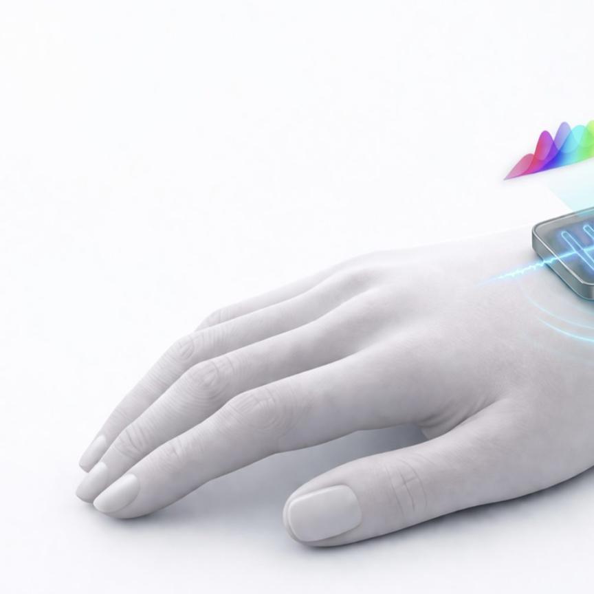
Interferometry in the palm of your hand
Miniaturised infrared spectroscopy technology, such as Hamamatsu’s MEMS FT-IR engine, is set to benefit many applications
Infrared spectroscopy is a versatile tool for numerous tasks such as process analysis, sorting, healthcare and food inspection. Since infrared light penetrates into materials, the composition of the material itself is observed instead of measuring only the surface.
This is possible thanks to the fact that molecular bindings are always in a state of oscillation. The infrared light interacts with those bindings and gets them to an excited state. The energy levels – and thus the amount of energy needed for such a transition – are unique for the respective atoms of the molecule, meaning only light of certain wavelengths is absorbed. By looking at the spectrum, that is the intensities of light plotted over the wavelength, samples can be analysed qualitatively and quantitatively.
As infrared spectroscopy does not require sample preparation and measurements only take about one second, it is easy to use for the operator. Furthermore, being a contactless and non-destructive technique, IR spectroscopy is low-maintenance, robust and hygienic. This allows accurate measurements while the sample is preserved and still usable.
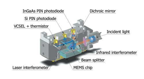
How FT-IR works
An important and widespread technique for wavelength dependent IR measurement is Fourier transform infrared (FT-IR) spectroscopy. A Michelson interferometer is one approach to distinguish the light intensity at different wavelengths: a dichroic mirror divides the light, one part of it is directed to a fixed mirror, the other part to a movable one. Both beams are reflected back to the beam splitter where they interfere with each other on their way towards an indium gallium arsenide (InGaAs) photodiode. As the movable mirror oscillates, its position is monitored with a semiconductor laser. This information and the interference pattern measured by the diode is finally translated into a spectrum using Fourier transform. Hence, the name Fourier transform infrared spectroscopy.
Why FT-IR saves resources
The compound material InGaAs, especially extended InGaAs with sensitivity up to 2.5µm, is a challenging raw material to grow due to a lattice mismatch between the optically active InGaAs and the substrate which can lead to crystal defects. Due to this, the smaller the active area required for the detector, the higher the production yield per wafer and the lower the material cost. As the FT-IR engine merely requires an InGaAs diode, it can be more cost effective than diffraction spectrometers with comparable characteristics.
Applications for FT-IR
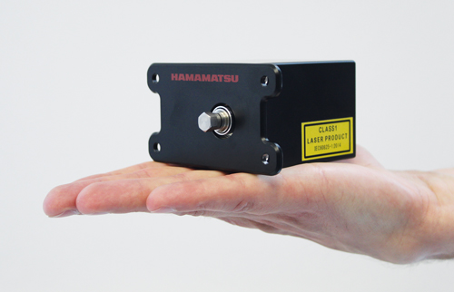
The optical interferometer is the biggest part of such a device, making most FT-IR spectrometers bulky bench-top instruments. By using our own unique MEMS technology, Hamamatsu has reduced the size so the whole device fits on a palm, enabling interferometry within handheld systems.
In order to achieve optimum optical characteristics despite minaturisation, a large 3mm dia. movable mirror ensures high sensitivity up to 2500nm, equivalent to tabletop devices. Additionally, the outstanding resolution and the great signal-to-noise ratio ensure that the FT-IR engine is suitable for sophisticated tasks such as:
•Environmental monitoring in the field;
•Ingredient analysis of agricultural products;
•Quality control in factory production;
•Food or plastic sorting at the store; and
•Point-of-care testing for human health at home.
Hamamatsu Photonics is a leading supplier of cutting-edge photonics technologies with more than half a century of experience. We are happy to help you design your sophisticated device. Please feel free to contact us and tell us more about your requirements.