With its non-destructive properties and resistance to water interference, the benefits of using Raman spectroscopy for analysis are widely recognised in the fields of life sciences and pharmaceuticals. The technique has great potential in the field of medicine but, as yet, has not been adopted widely in a medical field.
Hilde Jans, technical coordinator at research and development hub, Imec, explained: ‘We see advantages [of Raman] in endoscopy, but it is all still at the research phase. That is always the question, the chicken-egg problem. What the medical fields want to see is a proven relationship between the Raman spectrum, for example, and a disease. But people also have to implement that technology – so it’s always that balance.’
In the more cosmetic market, there have been greater strides commercially, said Jans. ‘There are tools on the market for checking the skin, for example, and so dermatology and skin profiling in general are very hot topics for Raman spectroscopy.’
The greatest advantage of Raman compared to other spectroscopic techniques in biological applications, explained Jans, is that there is no water interference. ‘Water overwhelms the spectrum in spectroscopy,’ she stated, ‘whereas it doesn’t do that in Raman spectroscopy because it is shifted. That is a huge advantage of using Raman in these applications.’
Is Raman spectroscopy too expensive?
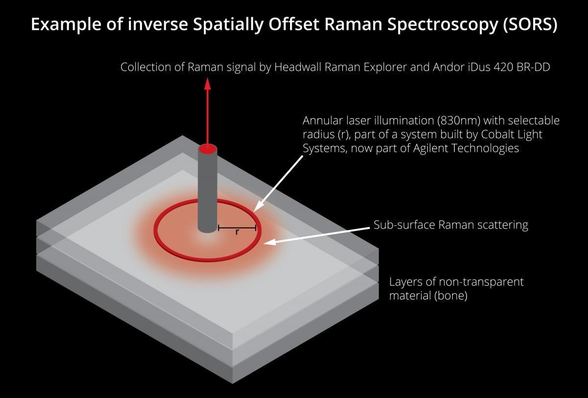
The perceived prohibitive cost of using Raman is one factor that could be limiting its use in many fields, Jans added. ‘A Raman spectrometer is a very expensive tool,’ she explained. ‘If you want to use a whole spatially offset Raman spectroscopy (SORS) system, it would easily cost £300,000 to your home, so that’s maybe a reason why it hasn’t been adopted that much yet in the medical space.’
One area of development across both research and commercial organisations that is hoped to counteract this, is reducing the size of the system. Jans continued: ‘Now there is lots of traction, so people are developing hand-held tools and even smaller devices – you really see the trend. I checked and there are almost 30 companies with several hand-held devices, but there is also the trade-off between the performance of these instruments and their size. We want to overcome this by building a very small form factor Raman spectrometer, which gives the same performance as the full size, and this is what Imec intends to do.’
The spectrometer – which was demonstrated at SPIE Photonics West 2019 in San Francisco – is an on-chip solution measuring 2cm in width and 500µm in thickness, and based on a newly patented concept for the company. Integrating a parallelisation of some 700,000 waveguide interferometers monolithically on top of a CMOS image sensor, both high optical throughput and high spectral resolution can be reached in a significantly miniaturised device. The system is built on Imec’s new silicon nitrate photonics (SiN) bio photonics platform and is compatible with high-volume manufacturing.
Imec's Raman spectrometer on-chip solution measures 2cm in width and 500μm in thickness
Application opportunities for Raman spectroscopy
A portion of the work was completed under the EU-funded IoSense programme, which aims to boost the competitiveness of electronic components and systems in Europe, and ultimately improve the time to market. Speaking of the potential applications Pol Van Dorpe, principal member of the technical staff at Imec, said: ‘With the right partners, we see many application opportunities in areas like food analysis, melanoma detection, or skin hydration. In the medical domain, we see opportunities for in-line measurements during surgery or endoscopy.’
Jans elaborated: ‘What we have new now is a SiN photonics platform. Silicon nitrate is the material that guides the light. We use plasma enhanced chemical vapour deposition (PECVD) technology for this, as it is a low temperature method to deposit these photonic layers, and we can do that on top of a CMOS imager. It is a smart design, so the system architecture – how you put this light and how you make the interferences – that is the trick.’
The spectrometer is currently at the prototype stage, and the company is undertaking feasibility studies to prove its effectiveness. ‘We have a roadmap as well,’ said Jans, ‘so at the end of the year we will have a small hand-held or benchtop device to demonstrate performance, but of course Imec is not a product builder. So, we are looking for partnerships for specific oriented applications that we can work on further and develop an application-specific product.’
Scaling down a benchtop Raman imaging system
Instrument maker Renishaw has also been developing its Raman technology to scale it down in size. Its current solution – a compact benchtop Raman imaging system designed for biological and clinical research – enables the identification and assessment of biochemical changes associated with disease formation and progression. It has the potential to aid surgeons in tailoring their surgical strategy based on a patient’s specific tumour genetics. Users are able to gather detailed information from biological samples, ranging from the distribution of exogenous and endogenous compounds within tissue, to the detection of protein secondary structure changes that arise due to drug interaction and tissue injury.
The benchtop device has undergone tests at numerous locations including the neuro-oncology department at Oxford Radcliffe Hospital (UK), where it was used to demonstrate discrimination between diseased and healthy brain tissue, studying tissue for the genetic classification of glioma tumours.
The spectrometer’s portability allowed for installation directly where surgery was taking place, so testing could be undertaken on samples immediately post-removal. It allowed for the building and testing of glioma tumour classification models, discriminating the cancer subtypes, in a number of tissue and cell types, and identifying biochemical changes associated with cancer formation and progression. The company plans to further downsize, according to senior application scientist Martin Isabelle, with the development of a fibre-optic Raman probe that could be used to gain surgical information in real time. However, it will first need to overcome the challenge of avoiding the reduction of an already relatively weak Raman signal, therefore rendering it un-useable.
Immediate results
Working towards commercialisation is Headwall Photonics, and the company likewise believes that the way forward in medical applications is to be able to offer OEMs smaller instruments, that deliver precise measurement accuracy. Christian Felsheim, business development and OEM account manager, firmly believes in Raman’s potential within medicine.
Raman is only one of the techniques used by the company, but Felsheim explained that it offers the unique benefit of providing information on both the atomic and also the molecular level. ‘That makes it officially interesting for any kind of life science for biological or medical application,’ he explained. ‘Most of the material is made from carbon hydron atomic material which, if you get that into other spectroscopy technologies, you find that out there is a lot of carbon, a lot of hydrogen, but you don’t know anything about the biological reference. Raman does that as it utilises the molecular structure to give you a signal you can record to identify and analyse the material. That’s what we have seen over the past decades. Raman spectroscopy has been used more in life sciences to get information about different materials.’
The challenge, said Felsheim, lies in the basics of Raman as it relies on the non-elastic scattering of light from the molecular structure – therefore giving, by design, a very weak signal. ‘In other applications such as machine vision, where you are working with dead materials, you solve the problem of low signal-to-noise by applying a huge amount of signal – a huge amount of light,’ he said. ‘You can’t do that in a biological probe because you would simply burn it. You need to be very sensitive about the amount of energy you are giving on your probe. With low light applications this would definitely be the case with Raman spectroscopy. You need very good, precise optics that really take advantage of every single photon coming back without scattering it.’
Approval process
In turn, the low signal-to-noise ration presents a logistical obstacle when it comes to commercialisation. Felsheim continued: ‘The challenge, especially in medical applications, is all the work you have to do to get this certified. Getting it approved, getting it through FDA, getting it through the medical approval in Europe, and CE approval. As you have a low signal-to-noise, you need more expensive equipment that makes the commercial side difficult for market entry.’
However, this is a question of development moving forward, he acknowledged. ‘The more the optical part is improved the more the sensor side is being improved,’ he said. ‘What also becomes more and more important is the software. Especially in this analytical area, we’re seeing that neural networks are being used more and more to do classifications of the signal coming back. We are moving towards that.’
For Headwall, the required solutions have been varied, so there isn’t one specific Raman solution in the pipeline for medical applications. Felsheim explained: ‘As it comes to these photon-starved applications where you only have a few photons over a very low signal, you must be even more precise that you project every single photon correctly according to the wavelength. That’s the strength of the technology, and we can customise these gratings to the specific need of the customer because they all need a slightly different spectral range,’ he said.
‘The nice advantage of using light as a measurement tool is it is non-destructive and that makes it applicable into medical applications, you just shine light onto exposed brain in a brain cancer operation without doing harm. You don’t need to cut out a probe; this non-destructive approach, with an immediate result, to me that’s the fascinating beauty of this technology,’ Felsheim concluded.
Glucose monitoring
One defined medical application that solutions are being developed for specifically is monitoring glucose levels in diabetes patients. A non-invasive spectroscopic device for this has been found to be just as effective as invasive techniques by an evaluative study conducted by researchers from the University of Missouri School of Medicine and the Massachusetts Institute of Technology (MIT). The results were recently published online by Analytical and Bioanalytical Chemistry.
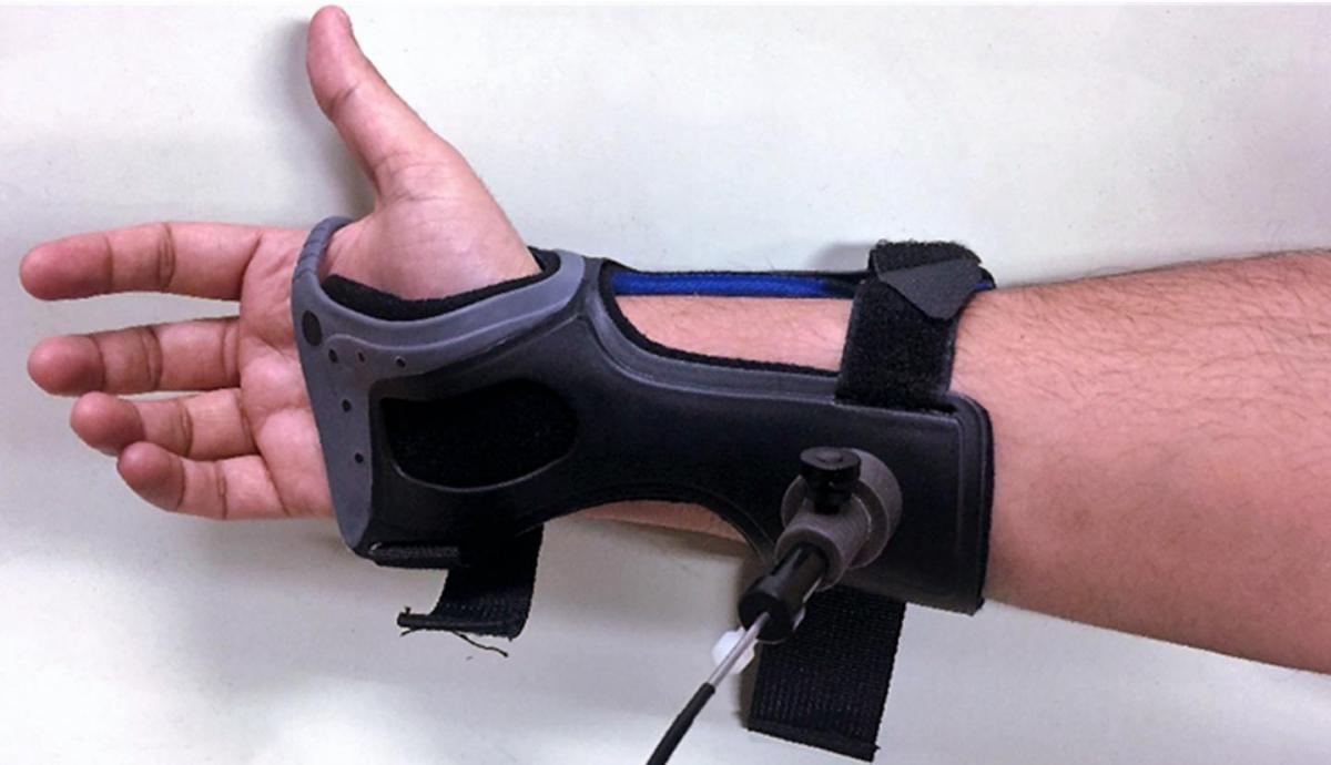
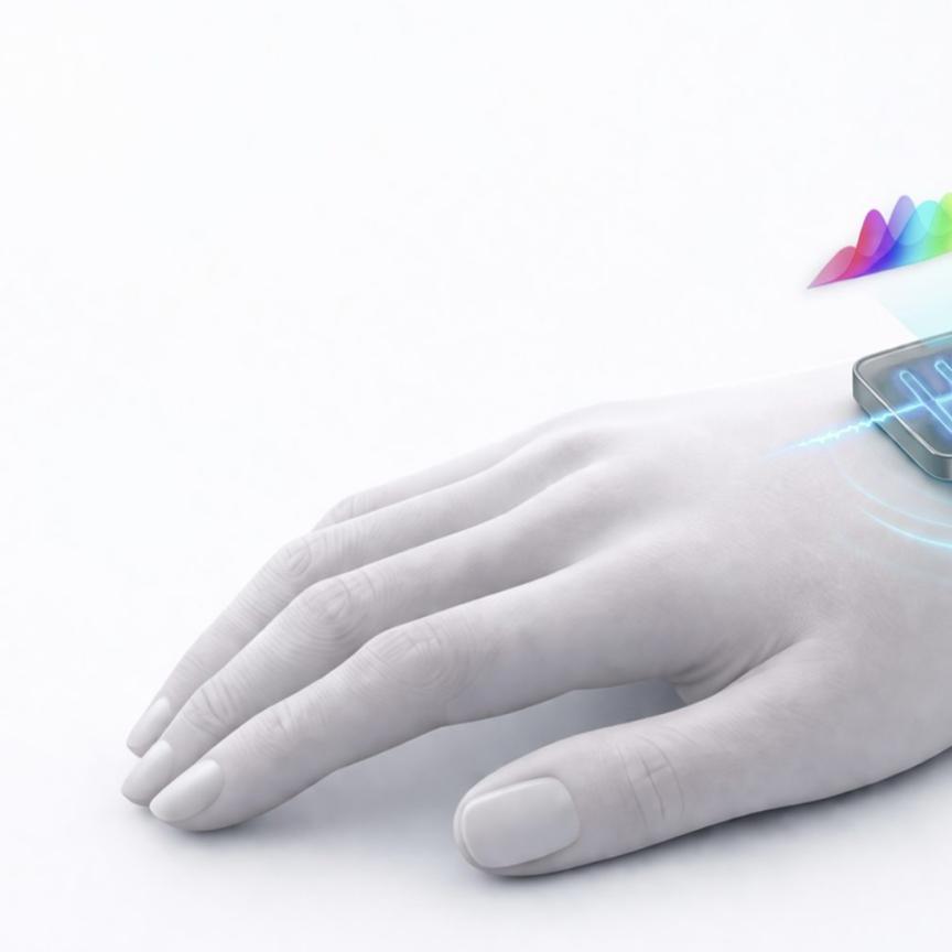
Raman spectroscopy blood-glucose monitor. Credit: Massachusetts Institute of Technology
Diabetics usually test blood glucose by pricking a finger, but this could soon be unnecessary thanks to a new device developed at MIT. The Raman spectrometer can measure the chemical composition of skin and determine blood glucose levels without collecting a blood sample. ‘With diabetes on the rise, the development of an accurate, efficient and inexpensive alternative method to test blood glucose levels is an urgent clinical need,’ said Dr Anandhi Upendran, director of biomedical innovations at the MU School of Medicine Institute for Clinical and Translational Science and co-author of the recent study.
The device delivers laser light via a fibre-optic cable to a wristband worn by the person being tested. The spectrometer can detect fat, protein, collagen and glucose molecules in the skin. The glucose present in the blood causes a shift in wavelength in the laser light that can be detected and used to measure glucose levels.
In the study, ‘Evaluation of accuracy dependence of Raman spectroscopic models on the ratio of calibration and validation points for non-invasive glucose sensing’, the researchers measured blood-glucose levels of 20 healthy, non-diabetic adults prior to taking a glucose-rich drink. Blood glucose levels were then measured at intervals over the next 160 minutes using three methods: spectroscopy via the new wristband, an IV blood test, and a finger prick. The spectrometer measured glucose values as accurately as the finger-prick test.
Commercial organisations are involved in this application. Wasatch Photonics has provided customised Raman solutions to researchers developing home-use non-invasive glucose monitoring devices. In a study involving 35 patients, which was published last May in Plos One, measurement performance for glucose levels present at, or below, a depth of ~250µm beneath the skin surface was comparable to that reported for currently available invasive continuous glucose monitors.