Ultrasound is a well-established imaging technique that has become standard in most clinics and hospitals. Now, more than 100 research groups are focusing on photoacoustic imaging – a hybrid of ultrasound and optical imaging that allows for deeper tissue depths with greater resolution – in an attempt to get the technique into clinics and hospitals.
It is estimated that photoacoustic imaging is still at least five years away from becoming common practice in hospitals and clinics. The imaging modality is still largely in the pre-clinical stage, with only one company currently having received CE marking for its photoacoustic system. This, in part, can be attributed to the high price of the technology, representing a demand for low-cost light sources optimised for photoacoustic imaging.
However, developments made in the last few years by both medical equipment manufacturers and research groups could assist the adoption of photoacoustic imaging in the medical sector. Such advances include the commercial launch of a photoacoustic instrument that can be attached to existing ultrasound systems, making clinical adoption easier, and several research groups are exploring cheaper alternative light sources for use in photoacoustic systems.
Attaching light
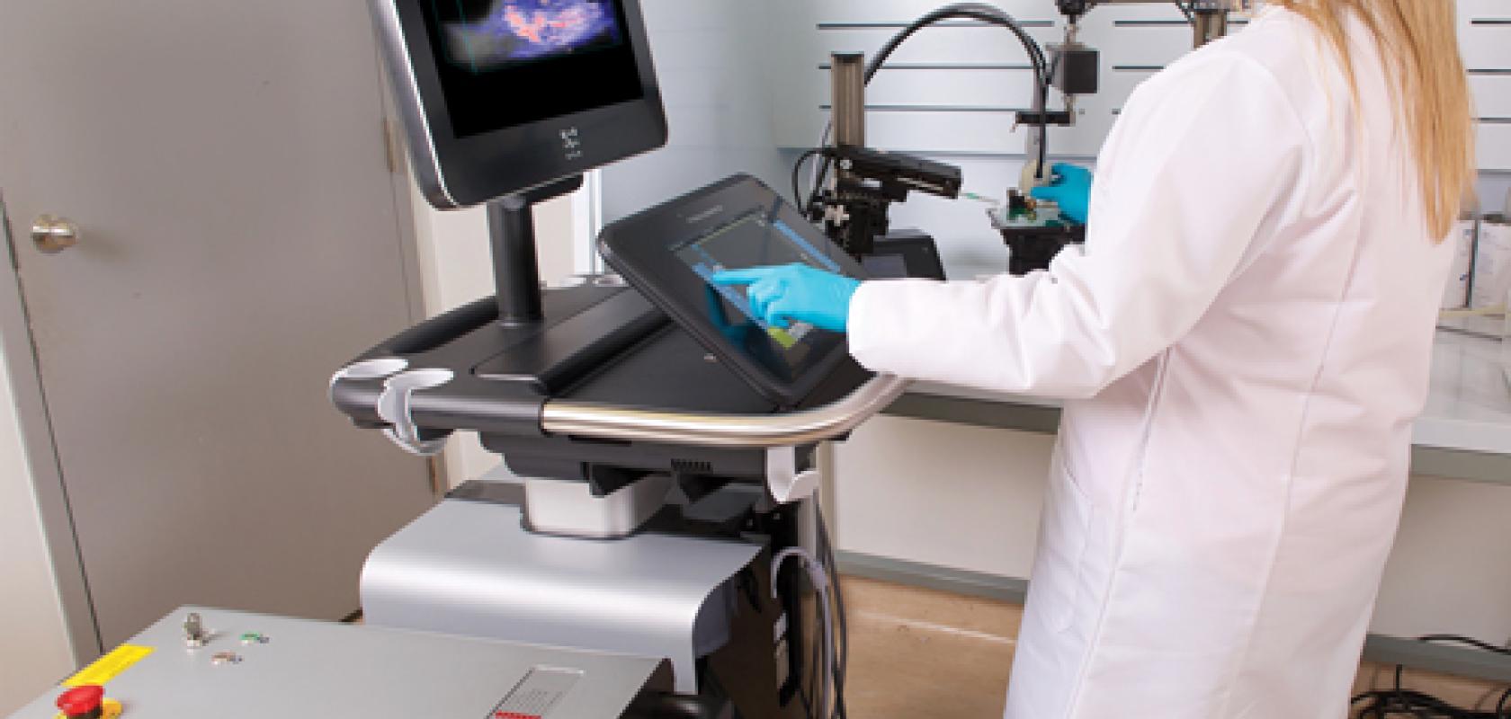
Fujifilm VisualSonics, a manufacturer of micro-imaging systems, offers its photoacoustic technology as a modal attachment to a pre-existing ultrasound system. ‘This will be an advantage in the future for the clinical applications in hospitals, where everybody uses ultrasound,’ explained Dr Jithin Jose, business development manager at Fujifilm VisualSonics. ‘People won’t have to use new technology or a new system. They can continue to use the same ultrasound system that we originally supplied with this new modality attached.’
The Fujifilm VisualSonics’ Vevo LAZR system is a hybrid imaging modality that uses an Nd:YAG laser, operating at wavelengths between 680-970nm, the range optimal for photoacoustic imaging. The laser light is coupled into fibres that connect to a handheld transducer, allowing the system user to move the laser light across the area of tissue being imaged.
When the tissue absorbs the laser light it undergoes thermo-elastic expansion, generating ultrasonic pressure waves which are then detected by 256 piezoelectric elements in the transducer head. The received information is then converted into high-resolution images that provide additional anatomical, functional and molecular information of the tissue that could only otherwise be obtained with other expensive imaging modalities, such as MRI or CT scanning. Additionally, unlike an MRI or CT scan, the approach is fast, non-invasive, non-ionising and less expensive.
Last month, Fujifilm VisualSonics released a new version of its photoacoustic imaging system, the Vevo LAZR-X, which operates at additional wavelengths (680-970nm and 1,200-2,000nm), allowing a wider variety of tissues to be imaged. The usability of the system has also been improved, with the addition of a touch screen interface. Usability and capability improvements such as these could lead to the photoacoustic imaging being adopted at a faster rate, as ultimately less training will be needed to use the more capable technology.
Although Fujifilm VisualSonics focuses mainly on pre-clinical imaging applications in oncology, neurobiology, cardiology and molecular imaging, it is also promoting further adoption of photoacoustics in hospitals. ‘We have started clinical trials in Europe looking into peripheral arterial diseases, breast imaging, wound healing and skin cancer,’ explained Dr Jose. ‘These are at really early stages at the moment, but the initial results are promising.’
Breast cancer imaging is one of the main intended uses of photoacoustic imaging, according to Dr Jose. ‘X-Ray (mammogram) or MRI imaging methods are currently used for this, but these are normally expensive and ionising technologies,’ he said. ‘With photoacoustics you can have a quick, non-invasive scan that provides a better understanding of the tumours.’
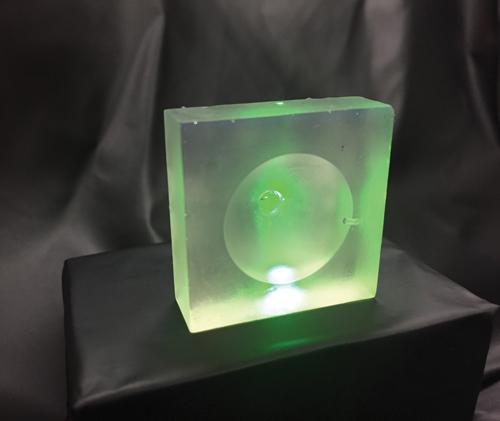
NTU's unique 3D printed resin lens overcomes the limitations of glass
Companies such as Canon currently have photoacoustic systems under development for this purpose, and although still in clinical trials, it is believed they will be used in hospitals within five to 10 years.
Right now, companies such as Canon are developing photoacoustic imaging devices for breast cancer imaging, according to Terence Wong of Washington University in St Louis. ‘Many people are working on [photoacoustic imaging] but it still needs some time. I believe that in around five to 10 years the breast scanner will become available in hospitals.’
Cutting costs
While introducing photoacoustics as an add-on to ultrasound could increase the ease with which clinics can adopt the technique, the high price of the lasers used in the systems could continue to deter potential users.
Research conducted by Wong at Washington University in St Louis has produced hopeful results that could reduce the cost of photoacoustic imaging dramatically. In research published in the Journal of Biomedical Optics, Wong proposed the use of xenon flash lamps as a cheaper alternative illumination source for photoacoustic imaging.
In the paper1, the researchers noted that the high purchase price and maintenance costs, as well as the bulkiness, of lasers, has hindered the rapid clinical dissemination of photoacoustic imaging systems.
‘One of the main reasons why hospitals are not taking photoacoustics into their clinics is because the laser in the system is very expensive,’ explained Wong. ‘If you can achieve the same performance at a much cheaper cost you can definitely increase the chance of persuading hospitals to use the systems.’
Photoacoustic imaging uses light sources of short pulse width and high energy to induce the ultrasonic waves. Xenon flash lamps match these criteria and can therefore provide a smaller, less expensive and lower maintenance alternative to lasers. In addition, it allows the whole system to be reduced in size, making it more portable. ‘The low cost and portability will facilitate the adoption of photoacoustic imaging from different perspectives in clinics globally,’ affirmed Wong.
In the published work, Wong demonstrated that the imaging quality achievable by lasers and flash lamps is very comparable. ‘Lasers are too good to be the light source in photoacoustic imaging for most situations – we don’t need the light to be well confined in a collimated beam for photoacoustic imaging, so we are really lowering the cost while maintaining the image quality,’ he explained. ‘This is why it has the potential to extend the usage of photoacoustic imaging, for example for global healthcare and in developing countries.’
Xenon flash lamps perform similarly to lasers in triggering photoacoustic waves; however, they have the further advantage of being able to provide a broader spectrum of wavelengths (200-2,000nm) than the standard Nd:YAG lasers currently used. At different wavelengths, tissue components absorb light differently and result in varying imaging contrasts. Using a broader spectrum therefore ensures that the optimal wavelength can always be used to achieve the best contrast.
Wong envisions that an eventual application of flash lamps in photoacoustic imaging will be skin melanoma imaging, preventing the need for invasive biopsies to study cancerous tissue. Wong and his colleagues have previously demonstrated this application of photoacoustics using lasers. The team were able to obtain enough accuracy in size, depth and volume information to allow a surgeon to determine a definitive treatment and prognosis. The next step is to achieve the same results using a flash lamp. ‘It would be a very big deal if we could do this because it would be a very cheap device that can check what surgery needs to be done on a skin melanoma.’
A reduction in price will not only make the initial purchase of a photoacoustic system more manageable, but will also help the future maintenance of the equipment. For example, during clinical trials, back up equipment is often required in case components fail and need replacing. ‘For lasers this is really hard to do, due to their cost, but it can easily be done with xenon flash lamps, which will really help facilitate these trials,’ said Wong. Flash lamps could also be integrated into existing clinical trials without difficulty. ‘The xenon flash lamp is just replacing the laser, so it would be a really cheap add-on for the system,’ confirmed Wong.
Portable photoacoustics
Although Nd:YAG lasers are currently the standard illumination source for photoacoustic imaging, the need for a powerful pump source creates a demand for water cooling, which can greatly increase the footprint (systems typically weigh between 50kg and 200kg) as well as the cost of the photoacoustic system.
‘That’s where we need more development for photoacoustics. The laser needs to be made as portable as possible,’ commented Dr Jose of Fujifilm VisualSonics. ‘This is where xenon flash lamps and laser diode based photoacoustic imaging is interesting.’
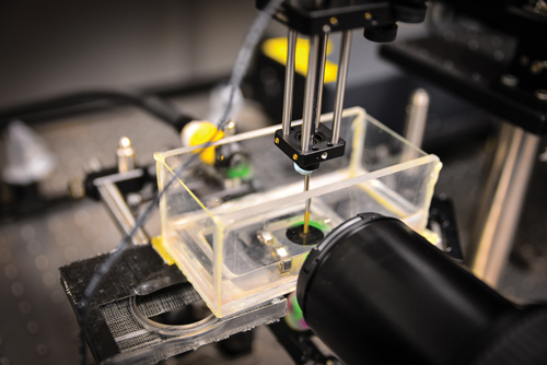
NTU's new ultrasound device fires ten pulses per second through the 3D printed lens, generating enhanced ultrasound or photoacoustic waves
‘I really like the idea of using xenon flash lamps, as the advantages of that will be, if it’s practical, being able to use all the wavelengths,’ Dr Jose continued.
He went on to explain a current need for dedicated photonics development for photoacoustics, particularly concerning laser diodes. ‘There is a lot of research going on in this area, but it is still in the early stage,’ he said.
Such research has recently been carried out at the University at Buffalo in America and published in Biomedical Optical Express. The authors had previously explored LEDs, laser diodes and diode-pumped solid-state lasers (DPSSL) as alternative illuminations sources in photoacoustic imaging. In this more recent paper2 published in 2017, the researchers demonstrate a compact diode-pumped laser, weighing only 1.6kg, being used to provide high-quality images at a depth of over 4cm in a portable photoacoustic system.
It was concluded that compact lasers based on emerging diode technologies are well-suited for preclinical and clinical photoacoustic computed tomography.
Meanwhile, an EU-funded project, called ‘Fullphase’ and involving a collaboration between 11 photonics partners, is developing a portable, multi-wavelength photoacoustic medical system using laser diodes. The system could apply to point-of-care early disease detection or optimal treatment monitoring in the fields of oncology, rheumatology and cardiology.
Dr Jose outlined the elements of photonics he believes need to be developed in order to improve the functionality of photoacoustic instruments and ultimately lead to increased adoption in medical facilities. ‘Anything to improve the repetition rates from 20Hz into the kilohertz range while maintaining high energies would be really ideal,’ he explained. ‘If we can achieve a broad range of wavelengths with a fast, high energy and compact system, then that will open up more photoacoustics applications.’
Stretching it further
The number of application areas of photoacoustic imaging is indeed a significant factor that will influence its clinical adoption. In another area of research being conducted at Washington University in St Louis, Pengfei Hai is using photoacoustic imaging to quantify the elasticity of tissues, an application previously unexplored by the technique. The results, which have been published in the Journal of Biomedical Optics3, indicate the potential for using photoacoustic imaging for assessing tissue elasticity for both research and clinical purposes.
A tuneable optical parametric oscillator laser pumped by an Nd:YAG laser was used by Hai and his colleagues. The photoacoustic system – which can measure the strain present in tissues – was used in parallel with a customised compression system that can measure stress. Obtaining these two quantities enabled an elastogram to be calculated for the tissues being examined.
‘We can map tissue elasticity on an absolute scale. It enables long term monitoring of tissue elasticity,’ Hai said. ‘It has many potential clinical applications, such as monitoring cervical elasticity, an important indicator of early birth during pregnancy.’
Hai and his colleagues are now working on a number of clinical trials, looking to find the best applications for photoacoustic imaging. ‘This technique can really be applied to help a large population… we are in the clinical trial phases and we are very confident and look forward to seeing the best clinical applications of photoacoustic imaging that make it widely adopted in clinics.’
With regards to melanoma tumour imaging, according to Hai, photoacoustic imaging ‘may eliminate the process of biopsy for the final diagnosis decision to be made,’ he said. ‘This is why the doctors are so excited about the technique.’
Before photoacoustic imaging can become the standard in melanoma tumour detection and be widely adopted in clinics, it has to be proven capable of replacing biopsies, and this calls for more trials to be carried out.
In working on multiple collaborative projects with doctors, Hai has discovered that, while some are very aware of the technology, others are not at all. He emphasised that further public awareness of photoacoustic imaging is needed in order to increase the adoption of the technique. In addition, Hai believes that coupling photoacoustics to ultrasound instruments – as is being done by Fujifilm VisualSonics – provides a ‘very natural entry point to the clinics for photoacoustic imaging, because photoacoustic and ultrasound imaging can share the same ultrasonic transducer, and ultrasound imaging is already used every day in clinics.’
References
[1] Wong, T. et al., 2016. Use of a single xenon flash lamp for photoacoustic computed tomography of multiple-centimetre-thick biological tissue ex vivo and a whole mouse body in vivo. Journal of Biomedical Optics 22 (4)
[2] Wang, D. et al. 2017. Deep tissue photoacoustic computed tomography with a fast and compact laser system. Biomedical Optical Express 8 (1) pp 112-123
[3] Hai, P. et al., 2016. Quantitative photoacoustic elastography in humans. Journal of Biomedical Optics 21(6)
At the Nanyang Technological University in Singapore, researchers led by associate professor Claus-Dieter Ohl have developed a device that generates enhanced photoacoustic waves using 3D printed lenses. By using additive manufacturing to produce the optics, the researchers were able to customise their lenses to best suit the tissues they work with, and to a degree of precision currently unachievable with glass.
‘The surface of a glass lens is not easy to change, the ranges of curvature are limited by the tools used to make them,’ explained Ohl. According to him, the acoustic properties of lenses are much more controllable with the plastic and resin materials used in 3D printing. In addition, moulding glass lenses is considerably more expensive compared to 3D printing them, which costs in the range of dollars and cents to make. Using additive manufacturing, the group at NTU has been able to produce lenses of optical quality equal to or even better than glass lenses, meaning a compromise in imaging quality does not have to be made when choosing 3D printing as a fabrication method for optics.
Ohl’s device differs from standard photoacoustic systems in that it is not used for imaging applications. Instead, the photoacoustic process is used to generate waves which then destroy or manipulate tissue. ‘On small scales, if you want to move cells to the left or right you can use these kinds of waves non-invasively,’ commented Ohl. ‘It produces very high frequency waves which are then focused to an area of around 100μm, where all of the energy is then delivered.’ As with standard photoacoustic systems, the device uses an Nd:YAG laser of a few nanoseconds in pulse duration, delivering tens of millijoules of energy to manipulate the tissues effectively.
Ophthalmic applications are currently being investigated by the group, as the lenses can be printed easily to the high level of precision required. A particular usage envisioned by Ohl would be the treatment of glaucoma. ‘The idea is to make a temporary incision in the eyeball to release the pressure, and we think very precisely focused photoacoustic waves could be a way to achieve that,’ he explained.
The technique also has potential to manipulate skin tumours, or deliver drugs to certain parts of the body, according to Ohl. As with standard photoacoustic systems, promoting a wider range of applications with the device will likely lead to an increased uptake of the photoacoustic technology.