Photoacoustic imaging has proven advantages over conventional imaging methods, but has not yet received clinical approval. According to Professor Jürgen Popp, speaking at the European Photonics Industry Consortium (EPIC) Biophotonics workshop in The Netherlands in November, if a biophotonic technology is to be useful not just in research but also in a clinical setting, then involvement within the scientific, medical and optics industries is fundamental.
Next year will witness a considerable movement towards a clinical translation: iThera Medical in Germany will start its first clinical trial on patients; and scientists from the University of Twente in The Netherlands will construct the first clinical prototype of their laboratory-proven photoacoustic mammoscope.
Photo- or opto-acoustic imaging uses a pulsed laser to illuminate the tissue being examined. When the cells absorb photons they heat up and expand. This excitation creates an ultrasonic pressure wave and it is this acoustic signal that photoacoustic imaging relies upon. ‘The absorption of the near-infrared (NIR) light at different wavelengths leads to the signal that we detect – this is the fundamental aspect of this type of imaging,’ said Wouter Driessen, probe development chemist at iThera Medical.
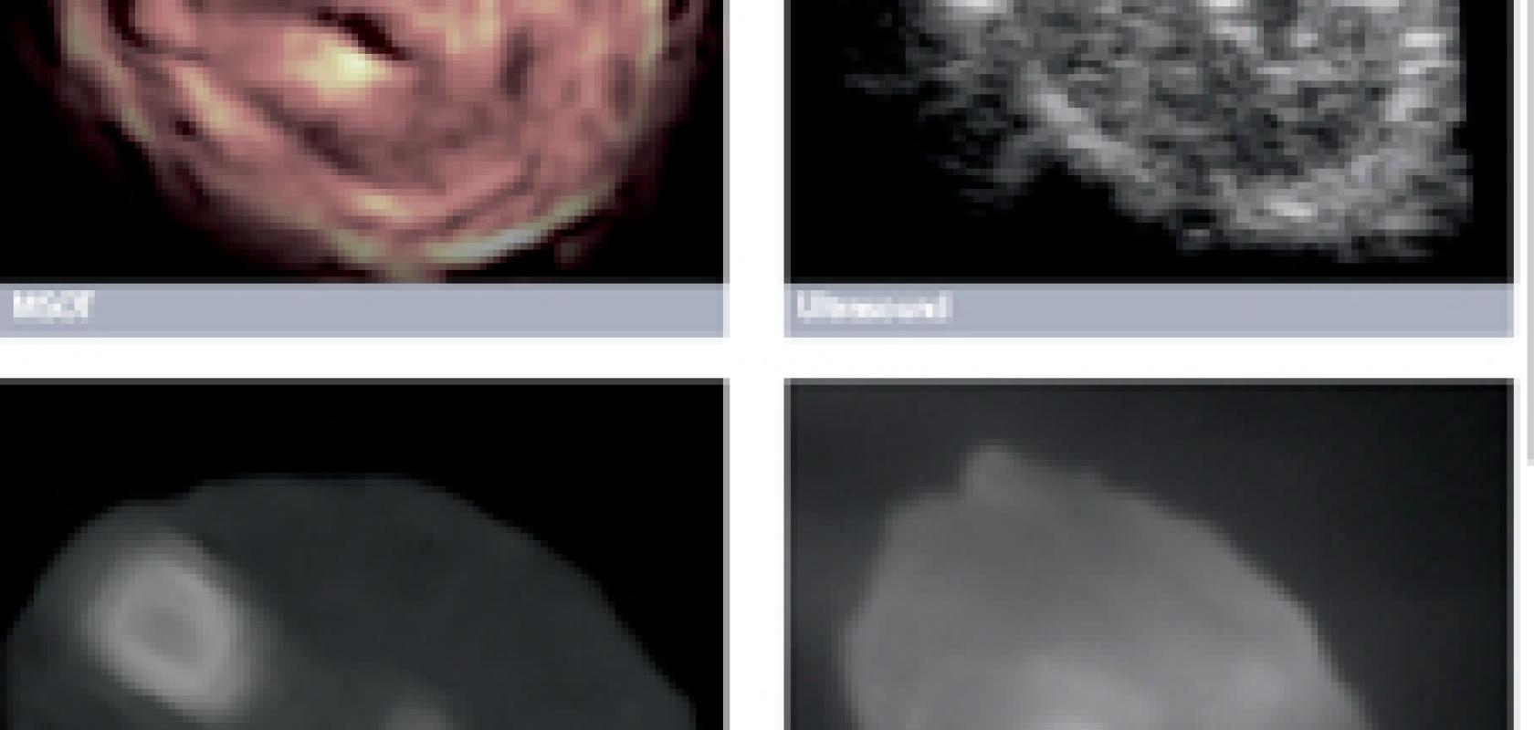
An optical contrast is created because different biological tissues have different absorption properties. ‘Something that does not absorb light in the NIR, such as water, will have a low level of absorption and emit a low photoacoustic signal,’ explained Christian Wiest, managing director at iThera. Alternatively, the tissue components that emit the strongest photoacoustic signals are: melanin, and the two main constituents of blood – oxy and deoxyhaemoglobin. ‘These three tissue chromophores have the strongest absorption and therefore dominate the intrinsic source of contrast in tissue,’ added Wiest. An ultrasound detector picks up the signal and iThera’s system converts it into a greyscale image to show vascular and tissue structure. It is also possible to un-mix specific signals spectrally, as Driessen described: ‘This allows us to operate more sensitively and specifically and show the doctor a heat map of the parameter of interest, such as oxygen saturation.’
Photoacoustic imaging combines the advantages of both optical imaging to offer haemoglobin-driven optical absorption contrast and ultrasound imaging to provide high resolution with a large imaging depth. ‘With this imaging technique, you can achieve the molecular specificity of optical imaging, combined with the depth and temporal resolution of ultrasound imaging,’ said Wiest.
Although this form of imaging is not yet licensed for human use, several organisations are working towards its translation into clinical settings. iThera Medical in Germany is marketing its Multispectral Optoacoustic Tomography (MSOT) scanners globally; these are intended for use on small animals in preclinical research, such as cancer imaging. In November, Driessen spoke at the BioNanoElectronics in ICT, Biomedicine and Development workshop in Finland to present pre-clinical results, which demonstrated how MSOT technology can be used to observe the oxygenation patterns of tumours without the use of contrast agents. He concluded his talk by expressing the hope that MSOT will soon be translated into the clinic.
iThera is already working in collaboration with academic institutions and pharmaceutical companies to complete small-scale trials with healthy volunteers. These showed that it is possible to perform real-time analysis of tissue chromophore distribution in humans. The next stage will be to begin trials on patients, which will include examining haemoglobin and oxygenation statuses in peripheral artery disease sufferers. ‘We are starting the first formally approved clinical trial this year, and are building a set of experimental imaging platforms that we will use in clinical settings,’ Wiest said. ‘It is expected that we will have CE-marked approval – which is equivalent to the FDA approval – in 12 to 18 months.’
There has already been a great deal of support for the translation of photoacoustic imaging into the clinic, explained Wiest: ‘What we’ve experienced so far is promising – we have seen a lot of our partners at university hospitals carrying out both small-animal imaging and also translation testing for humans,’ said Wiest. ‘Research groups are still investing and submitting protocols to test photoacoustic imaging on patients.’ Moreover, Wiest explained that there is an unusual pattern occurring in the development of this technology, which highlights its clinical potential: ‘Unlike other technologies where the vendor would need to pay physicians to carry out testing, it is the other way around – we are getting paid to support these studies. This shows that there is a lot of confidence in photoacoustic imaging to make a good clinical translation.’
Potentially, the clinical application of photoacoustic imaging will have a huge impact on the medical industry, according to Driessen: ‘The technology would be cheaper for hospitals than that used in other imaging modalities such as MRI and PET. For the patient, it will be more-or-less the same experience of an ultrasound – painless and relatively stress-free.’
However, the conversion into the clinic will not come without its challenges. iThera scanning systems are currently operated only by skilled users in controlled environments, such as research laboratories. To construct a system that is robust enough for day-to-day use in a hospital presents other difficulties, as Wiest described: ‘The laser technology is quite sophisticated and sensitive, so the technical challenges [are] to build something sufficiently sturdy for clinical use.’ iThera co-developed their existing MSOT machines as a result of a close collaboration with a laser partner based in Munich, Germany. It is evident that collaborations between research groups and the industry are needed to push photoacoustic technology towards commercialisation.
Professor Jürgen Popp, scientific director at the Institute of Photonic Technology (IPHT) in Germany, shares this view of the need for strong involvement from both the industry and healthcare professionals. His talk to the EPIC Biophotonics workshop at the end of November focused on ‘Optimising the Road to Clinical Products’. According to Popp: ‘Medical professionals are important, because they define the unmet medical need – if there is no real need, it will be very difficult to introduce new technologies into the medical market. In order for this to happen, there needs to be increased interaction between the scientists, the physicians (end-users) and the industry.’ Communication between the sectors also plays a vital role in aiding the development of the laser components to be able to meet medical standards. Laser companies will need to have a greater understanding of medical regulations, Popp implied: ‘It is not comparable to developing a new laser system for the automotive industry, for example. It is a completely different issue, because the medical field has very specialised requirements.’
The IPHT based in Jena, Germany has recently established a research campus that will, together with industry, develop optical/photonics devices for the rapid and easy detection of infectious diseases. Using this project as an example, Popp again drew attention to the fact that, in order for photoacoustic technology to become fully commercialised, there needs to be a strong involvement from all fields, starting with the initial research stage through to the end translation into industry. ‘This development at IPHT is a public-private partnership between industry and research institutes, as well as clinicians and end-users. This is something that is completely new – we are starting the research together with the industry, so at the end we will have a smoother translation into the medical market,’ added Popp. ‘The idea is to have collaboration with the industry from the very beginning.’
A research group that will rely on close collaborations with the optics industry in the next year is a team from the University of Twente in The Netherlands, which has developed a laboratory prototype system designed for 3D photoacoustic full breast tomography. The prototype was tested by using a breast phantom, and the results were published in October’s issue of The Optical Society’s journal, Biomedical Optics Express. The researchers expect to have a clinical prototype ready by next year which will be a step towards a clinical transition, and the assembly of the optical components will depend on existing and increased partnerships with optics and photonics companies.
The photoacoustic system consists of an imaging tank containing ultrasound detectors and optical fibres, which is rotated around the breast to obtain a number of measurements at different positions. A high energy pulsed laser is employed to illuminate the breast. ‘For photoacoustic imaging the laser needs to operate in the NIR – so a wavelength of between 680 and 980nm – but higher wavelengths are also suitable,’ said Driessen. The Twente research team chose an Nd:YAG pulsed laser with a longer wavelength of 1,064nm: ‘We needed to have a high-energy laser because the light fluence has to be spatially distributed on the breast phantom surface,’ explained Srirang Manohar, assistant professor at the University of Twente. The laser beam is spread out to cover the entire phantom surface by using a fibre bundle that has nine outputs, all of which have an active diameter of 3.5mm.
The output laser energy delivered by the fibre bundle is roughly 80mJ (9mJ out of each of the nine fibre outputs), which is below the maximum permissible exposure for near infrared light. However, 80mJ is the maximum output energy that the fibre bundle can tolerate; even though the laser has maximum output energy of approximately 300mJ.
For a future clinical model, the team believes that a combination of fibre bundles and free-beam light delivery will be required. ‘The existing fibre bundle was custom-made by LightGuideOptics in Germany,’ said Manohar. It is expected that a close collaboration will need to continue to develop and improve these optical parts.
In order to increase the diagnostic power of the photoacoustic mammoscope, the team decided to employ an additional excitation wavelength. ‘In previous trials we used a single wavelength of 1,064nm and obtained some excellent results with a high level of contrast. However, we had some questions because we were not completely sure of the source of the contrast from the tumour – “was it coming from haemoglobin, oxyhaemoglobin, water, lipids or collagen?”’
To overcome this, a second wavelength of 755nm was chosen because at this wavelength the absorbance from water and lipids is low. For the recently published work, the team used a dual laser system which pulses at 1,064 and 755nm wavelengths. Both wavelengths share the same optical output parts and so can be coupled to the same fibre bundle. The laser system can be operated either in a single-wavelength mode (755nm or 1,064nm) with a 10Hz repetition rate or in a double-wavelength mode interleaving the two wavelengths with a 20Hz repetition rate. ‘We approached Quanta System in Milan, Italy which provided us with the dual laser system. When we commercialise this mammography system we would like to continue our collaboration and use this company again,’ said Manohar
The published trial demonstrated the capability of photoacoustic tomography to visualise different-sized vasculature objects, down to a depth of 40mm in the breast, with a high level of sensitivity. ‘However, what we presented in our work is a laboratory version of the system. So, it is not yet in such a form as to be used on a human patient,’ said Manohar.
The team hopes to have a clinical prototype ready in 2014. ‘We are quite sure that for this photoacoustic system to become commercialised, we will have to increase our dealings with optics companies. There are two aspects: the laser, and the optical bundles for light delivery – both fronts will require close collaboration with the industry.’