Miniature microscopes that can be housed in the tip of an endoscope are now being designed to improve cancer diagnosis.
Two research groups, from the Fraunhofer Institute for Photonic Microsystems (IPMS), in Germany and from Stony Brook University in the USA, are taking advantage of micro-electro- and micro-opto-electro-mechanical systems (MEMS/MOEMS) technology to build tiny microscopes for in vivo diagnosis of suspected cancerous lesions.
The devices, which make use of MEMS mirrors less than a millimetre in size, are designed to bring the microscope to the tissue, and, in future, could replace existing lengthy laboratory-based procedures.
Diagnosing cancer usually involves a biopsy followed by tissue analysis, which is time-consuming. Histopathology, which is currently the gold standard for diagnosis, is carried out in a laboratory by a specialist pathologist, and involves cutting, staining and examining tissue samples under a microscope.
However, not all lesions turn out to be cancerous, and unneeded biopsies are not only painful and stressful for the patient, but are expensive for healthcare authorities.
Advances in MEMS technology over recent years has allowed the development of tiny microscopic devices that perform in a similar manner to large desktop laboratory machines. Dr Jonathon Liu, who chaired a session titled ‘Miniature instruments for endoscopic microscopy’ at the MOEMS-MEMS conference at Photonics West in February, said that the use of confocal microscopy in endoscopic devices was a theme that came up: ‘There were a few talks on the use of MEMS mirrors for scanning a laser beam in a confocal microscope. That’s the most common use of MEMS mirrors in endoscopy – to scan the illumination light into the tissue to create an image.’
A confocal microscope is designed such that out-of-focus light is eliminated to increase resolution and contrast, and to image objects clearly in low background light. ‘In order to do that, these technologies [confocal microscopy] typically only allow you to image a single pixel within a tissue at a certain time,’ said Dr Liu, assistant professor of the Biomedical Engineering department at Stony Brook University in New York, USA. ‘So, in order to create an image, you need to scan that spot through the tissue – so that you reconstruct the image point by point.’
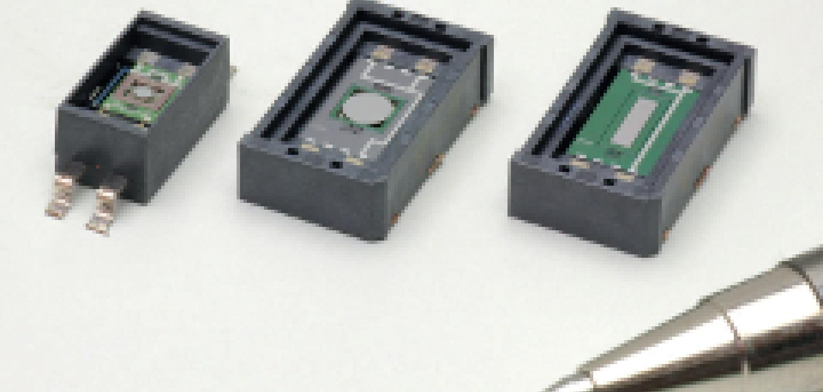
A team of scientists at the Fraunhofer Institute for Photonic Microsystems (IPMS) in Germany has developed a MEMS microscope head that can operate as a confocal system. The microscope uses a MEMS scanning mirror measuring less than a millimetre; the device itself has a diameter of just 6mm and is designed to fit on the tip of an endoscope.
‘The advantage [with the MEMS mirror] is that you can use a confocal system. Conventional scanners are way too big to be incorporated into the tip of an endoscope,’ said Dr Michael Scholles, head of business development and strategy at Fraunhofer IPMS. ‘We used a MEMS mirror with a diameter of a fraction of a millimetre so it then fits into the tip [of the endoscope].’
The endo-microscope can be part of a larger system for in vivo analysis of cells and other microscopic biological structures in real time, allowing doctors to diagnose cancer more rapidly.
The microscope head uses light from an external laser transmitted via an optical fibre. The MEMS scanning mirror fitted in endoscope deflects the laser beam and illuminates different points of the tissue as it oscillates. The light reflected by the tissue is detected by a sensor, and a two-dimensional image is reconstructed by combining the position of the mirror and image sensor signals.
The micro-mirror is able to oscillate in more than just one plane so that the laser beam can be reflected in any direction. This is achieved by a gimbal suspension which allows the scanning mirror to be deflected on two different and mutually independent levels, making it flexible and able to replace complex constructions usually employed to deflect laser beams.
The MEMS mirror was designed and manufactured at Fraunhofer IPMS on six-inch silicon wafers by photolithography. As many as 1,000 chips can be produced on a single six-inch wafer, which will be advantageous when the micro-endoscope goes into mass production, according to Scholles: ‘One advantage of producing MEMS devices on a wafer level is that if you go to higher quantities they become inexpensive – with one fabrication step you get a lot of individual chips.’
However, MEMS technology is being used in more and more new devices, and higher volumes are not always required in the development stage. In this situation, the process is more costly. ‘If you have a specialised application where you only need a handful of devices then you still have to pay for the whole lot [of MEMS components],’ explained Scholles. ‘So it’s a trade-off between the possibilities of higher-volume fabrication based on MEMS processes and the demand from the customer.’
A difficult part of the development was to produce a suitable micro-assembly for the endoscope head. ‘The technical challenge was the need to come up with the mounting technology for incorporating the MEMS mirror plus fixing the glass optical fibre,’ Scholles said. ‘Here we faced the challenge of making the complete system suitable for installation in the endoscope, and we managed to do it.’
Scholles went on to say that the integration of the MEMS component helped the researchers in other projects that are using a similar setup, such as a biometric device currently under development at Fraunhofer IPMS that scans the retina of the eye. ‘A lot of knowledge that we gained in the endoscope design has been carried over to the retina scanning system,’ Scholles added.
The IPMS team is currently searching for direct commercialisation of the device and is in talks with endoscope manufacturers.
Meanwhile, at the end of this year, scientists from Stony Brook University (SUNY) in New York, USA anticipate that their latest miniature handheld microscope will be ready for first-in-human studies. The microscope, which is also being incorporated into an endoscope, uses dual-axis confocal microscopy – a modified version of confocal microscopy that provides clearer images with better contrast. The device works in a comparable fashion to the Fraunhofer system, and employs a MEMs mirror to deflect a focused laser beam, transmitted via an optical fibre, into the tissue. As the MEMS mirror tilts in two dimensions, the system is able to scan the position of the beam and recreate an image in a point-by-point fashion.
One application the scientists are aiming for is the detection of oral cancer at an earlier stage, and they are hoping to approach the gold standard of pathology, according to Dr Liu at Stony Brook: ‘We are trying to see cells and sub-cellular structures on a microscopic level, in order to approach the gold standard of pathology. Instead of taking tissues out and placing them under a large microscope, we’ve decided to shrink down the microscope and bring it directly to the tissue.’
To image over a range of depths, one possible strategy is to use a MEMS mirror to deflect the laser beam further into the tissue. One aspect the SUNY team has been working on is a suitable way to move the MEMS chip to give the laser beam more penetration. ‘The MEMS mirror provides you with the tilting that scans a horizontal plane in the tissue,’ Liu described. ‘If you want to image deeper, which is in the vertical direction, you need to move the MEMS mirror forwards and backwards.’ To move the mirror, the team is using piezo actuators that are able to shift the MEMS component forwards or backwards by a couple of hundred microns. ‘So, the MEMS mirror is mounted on another stage that doesn’t use MEMS technology, but piezo technology, to push the MEMS mirror,’ said Liu. ‘We’ve also used small motors to do this.’
One shortcoming with microscopy as an imaging technique is that it is not able to measure beyond a depth of half a millimetre into tissue. But as the majority of cancers originate at epithelial surfaces, micro-endoscopes are well suited for their diagnosis. However, achieving a higher level of depth would provide more diagnostic information to the doctor, such as if the cancer has spread to underlying tissues. ‘One thing the clinicians would like to see is the depth of invasion,’ said Liu. ‘If the cancer has invaded into the deeper parts of the tissue it means that the cancer has metastasised to other parts of the body. We, and others, are working on innovative strategies to increase the image depth of these microscopes.’
Unlike Fraunhofer, which is producing the MEMS scanning mirror, the SUNY team has chosen to outsource from a commercial company in order to shorten the time to market. ‘We deliberately chose to use a commercial mirror from a start-up company in this case, because we wanted a reliable device that was already fully tested,’ Liu explained. ‘It would have been possible to do something new, and work with a collaborator to develop something with capabilities that aren’t available commercially. But that’s a trade-off – we wanted to build a device that we could get into the clinic.’ Mass production was also a factor to consider, as the University would not be able to produce the MEMS component in high volumes, according to Liu: ‘In our research lab, to scale up into a larger scale of manufacturing mode would be challenging. But these companies have mechanisms to produce large quantities of the mirror.’
And, this MEMS component was a central part of the design. ‘We designed our microscope around [the company’s] mirror. It was easier for us to design all the parts of our microscope, and to have them machined so that they could fit with the mirror which was already fabricated,’ Liu added. ‘The MEMS mirror is the most complicated part – it is the brain and the engine of the microscope.’
Another partnership that is combining the expertise in MEMS technology of one company with the mass production capabilities of another is that of Swiss company Lemoptix, which specialises in compact laser scanning systems and Japanese optoelectronics supplier Hamamatsu Photonics. The two companies are developing and commercialising devices based on MOEMS technology.
‘Lemoptix has been working in this field for more than 10 years and had already established the [MEMS] technology. Hamamatsu had the expertise and capabilities to scale it to volume production,’ said Craige Palmer, general sales manager at Hamamatsu Photonics UK. ‘Hamamatsu has taken that technology, industrialised it, and made it compatible with our own manufacturing facilities located in Hamamatsu City, Japan. We have scaled the technology up to volume production, required for emerging huge markets.’
According to Palmer, there is enormous potential for MOEMS devices in the medical industry: ‘The point about MOEMS devices is that you can make things smaller. In the medical market, the instruments used to be bulky or desktop. But now you can make mirrors smaller and lighter weight, so it’s possible to make portable versions of these instruments.’ Hamamatsu anticipates that the miniaturisation enabled by MEMS technology will give rise to the development of medical testing devices that patients will be able to use at home. ‘You will be able to tell what the problem is, without having to go to the hospital for a blood or urine test. So it is benefiting the consumer now,’ Palmer commented. ‘Whereas before, where Hamamatsu was selling to system integrators that developed products for hospitals, we can now envisage that kind of technology being brought into the home.’
Although MEMS scanning mirrors are becoming a mature technology, Dr Liu feels that in the biomedical field, until devices with MEMS mirrors are used as a standard of care in the clinic, the technology will not become mainstream: ‘The technologies are there, but we just need to find the right application that fully utilises these MEMS mirrors, and shows that the MEMS mirrors are the most ideal technology.’
Liu concluded: ‘Until we can demonstrate an ideal application that improves patient outcomes, it will be sort of a niche field – there will be a few groups utilising MEMS mirrors as part of their devices, but it’s not going to be a mainstream technology. That’s what we [at Stony Brook] are working towards.’
About the author
 Jessica Rowbury is a technical writer for Electro Optics, Imaging & Machine Vision Europe and Laser Systems Europe.
You can contact her on jess.rowbury@europascience.com or on +44 (0) 1223 275 476.
Find us on Twitter at @ElectroOptics, @IMVEurope, @LaserSystemsMag and @JessRowbury.

