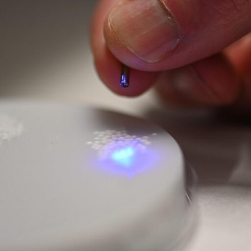
Scientists are building a new super-resolution microscope to study the pathogenicity of superbugs as part of a €5.6 million EU project.
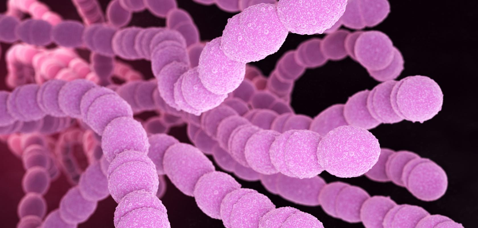
The microscope will allow scientists to observe bacteria like Streptococcus Pneumoniae at a resolution ten times higher than existing fluorescence microscopy techniques.
It is being developed as part of a project that has been awarded €5,635,529 by the European Commission via the Photonics Public Private Partnership.
The project is called 'Nano-scale visualisation to understand bacterial virulence and invasiveness based on fluorescence nanoscopy and vibratational microscopy (or ‘NanoVIB’ for short).
The project will allow scientists to observe how bacterial proteins localise on the surface of bacteria and study the interaction of the pathogen with immune and host cells.
A leading cause of bacterial pneumonia, meningitis, and sepsis, Streptococcus Pneumoniae bacteria are estimated to have caused around 335,000 deaths in children aged five years and under in 2015 worldwide. Current technologies do not allow a resolution that enables thorough studies of bacterial properties that affect disease development.
Ten-fold resolution
While super-resolution microscopes already exist, the NanoVIB team proposes to make a new device with unrivalled resolution capable of revealing the intricate, detailed molecular mechanisms underlying inter-and intracellular processes and disease.
Project coordinator, Professor Jerker Widengren, said: 'We expect our new microscope prototype to be a next-generation super-resolution system, making it possible to image cellular proteins marked with fluorescence emitters (fluorophores) with a ten-fold higher resolution than with any other fluorescence microscopy technique.'
With the help of advanced laser, detector and microscopy technologies that will be developed in the project, super-resolution localisation patterns of specific proteins will be overlaid with light-scattering images, correlating these patterns with local structures and chemical conditions in the bacteria.
Widengren explained: 'It works based on the so-called MINFLUX concept, where infrared laser light excites fluorophore-labelled molecules in a triangulated manner – leading to an increased resolution. The user can then fine-tune the microscopic imaging to previously unimaginable resolutions.
'MINFLUX microscopy will make it possible to resolve how certain pneumococcal surface proteins are distributed on the bacteria under different cell division stages, and whether these proteins are localised in such a way that specific, extra sensitive surface regions of the bacteria, a critical step of the cell division, are protected from immune activation.'
European Research Ecosystem
The NanoVIB team took their inspiration from a previous EU-funded project, Fluodiamon, which analysed how specific proteins are spatially distributed in breast and prostate cancer cells compared to those in corresponding non-cancer cells, demonstrating a new basis for cancer diagnosis.
The goal of the NanoVIB project is to retrieve information, which is not within reach by any other microscopic or photonics-based technique. 'We will demonstrate how cellular nanoscale protein localisation patterns can be resolved, which will help us reveal bacterial disease mechanisms and are likely to be of considerable relevance for many other diseases,' said Widengren.
'These studies could shed new light on how specific surface proteins of these bacteria are spatially distributed on the cells and provide important evidence that the virulence (capacity to generate disease) and invasiveness of these bacteria are strongly coupled to such spatial distribution patterns.'
The project will conclude in 2024 and includes six partners from three countries: Kungliga Tekniska Hoegskolan (KTH), the coordinator, Karolinska Institutet (Sweden); Institut für Nanophotonik , Abberior Instruments GMBH, APE Angewandte Physik und Elektronik (Germany); and Pi Imaging Technology (Switzerland).