Researchers have developed an ultrasound technique that enables optical images to be taken through biological tissue to image organs within the body, which they say could one day remove the need for invasive visual examinations using endoscopic cameras.
In Light: Science and Applications, Maysam Chamanzar and Matteo Giuseppe Scopelliti from Carnegie Mellon University in Pennsylvania, USA, showed they can use ultrasonic wave patterns to create a virtual ‘lens’ within the body, which can be used to ‘focus’ light within the tissue without needing to implant a physical lens. They say this enables them to capture images previously inaccessible through non-invasive means.
Biological tissue blocks most light, especially light in the visible range of the optical spectrum. Therefore, current optical imaging methods cannot use light to access deep tissue in the body from the surface. The researchers’ method, however, can be used to induce more transparency and enable more penetration of light through such tissue. This is because ultrasound waves can be used to compress and rarefy, or thin, whatever medium they are flowing through, which alters the speed that light travels through the medium – light travels more slowly in the compressed regions compared to the rarefied regions.
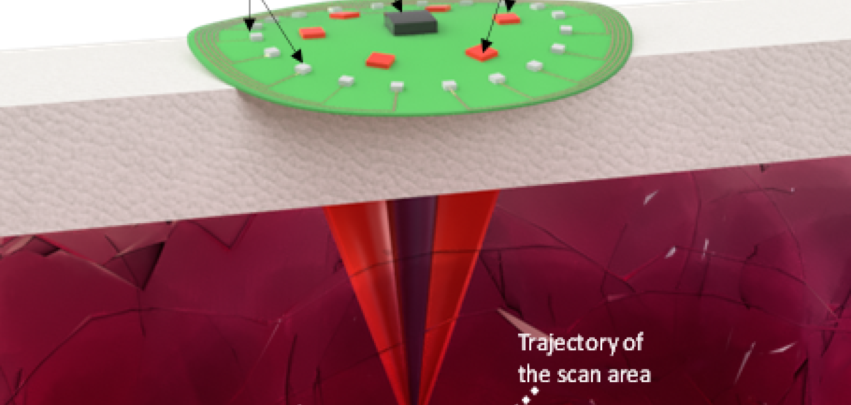
The team has shown that this compression and rarefication effect can be used to sculpt a virtual lens in biological tissue for optical imaging. By simply reconfiguring the ultrasound waves from outside, the virtual lens can be moved around without disturbing the medium. This enables imaging different target regions, all non-invasively.
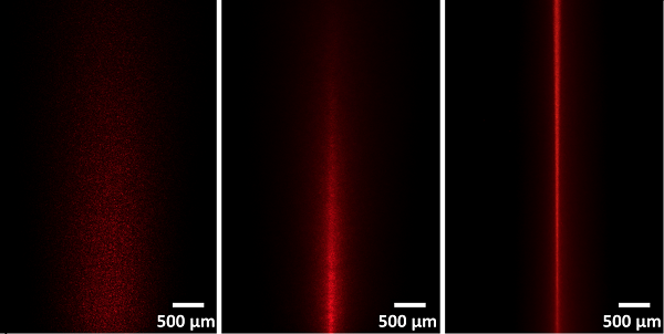
Left: Dispersion of a laser beam passing through tissue without ultrasound. Middle: Laser beam focused by ultrasound in a scattering tissue. Right: Laser beam focused by ultrasound in a minimally scattering tissue phantom. (Image: Carnegie Mellon University College of Engineering)
‘Being able to relay images from organs, such as the brain, without the need to insert physical optical components will provide an important alternative to implanting invasive endoscopes in the body,’ remarked Chamanzar. ‘This method can revolutionise the field of biomedical imaging.’
While the researcher’s paper is focused on the method’s efficacy for closer-to-the-surface applications, the team has yet to find the limit to how deep within the body’s tissue their ultrasonically-assisted optical imaging method can reach.
Putting it into practice
The published method could be applied in a number of different applications, according to the researchers. In the future, for example, it could be implemented in the form of a handheld device or wearable surface patch, depending on the organ being imaged. By placing the device or patch on the skin, the clinician would be able to easily receive optical information from within the tissue to create images of what’s inside, without the many discomforts and side effects that come with endoscopy.
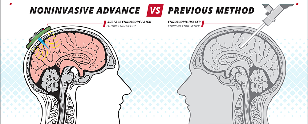
A noninvasive skin patch is one way the researchers’ ultrasound-based method could allow for easy imaging of the brain. (Image: Carnegie Mellon University College of Engineering)
The closest current applications for the new technique could be endoscopic imaging of brain tissue or imaging under the skin, but the researchers say the technique can also be used in other parts of the body for imaging. Potential clinical applications include diagnosing skin disease, monitoring brain activity, and diagnosis and photodynamic therapy for identifying and targeting malignant tumours.
Beyond biomedical applications, the researchers say the new technique could also be used for optical imaging in machine vision, metrology, and other industrial applications to enable non-destructive and steerable imaging of objects and structures at the micron scale.