A team of Polish researchers has developed an optical coherence technology (OCT) system capable of providing detailed images of the entire eye using electrically tunable lenses and a newly commercialised swept light source.
Published in Optica, the new device is capable of producing higher quality images than are currently possible, and could make eye examinations faster and more comfortable by removing the need to use multiple instruments when looking at different areas of the eye.
‘Diseases such as glaucoma affect both the front and back portions of the eye,’ said Ireneusz Grulkowski, whose research team at Nicolaus Copernicus University, Poland, worked with Pablo Artal’s team at the Universidad de Murcia, Spain to develop the new imaging system. ‘An instrument that can examine the whole eye will improve the patient’s experience because they won’t have to go through imaging with different devices. It might also one day reduce the number of instruments – which can be quite expensive – needed in an ophthalmology clinic.’
The researchers’ new OCT system is not only capable of imaging both the front and the back of the eye, but also the interfaces of the eye’s vitreous gel with the retina and lens in exceptional detail. This new imaging capability could allow scientists to better understand how the vitreous gel that fills the eye interacts with the retina and why it can sometimes become detached with aging.
‘We also want to use our instrument to measure opacities in the eye’s crystal lens and the vitreous to better understand how various parts of the eye affect the deterioration of vision,’ said Grulkowski. ‘We believe that the ability to measure these opacities and other properties of the eye that couldn’t be examined before will open up many new ophthalmology applications for OCT.’
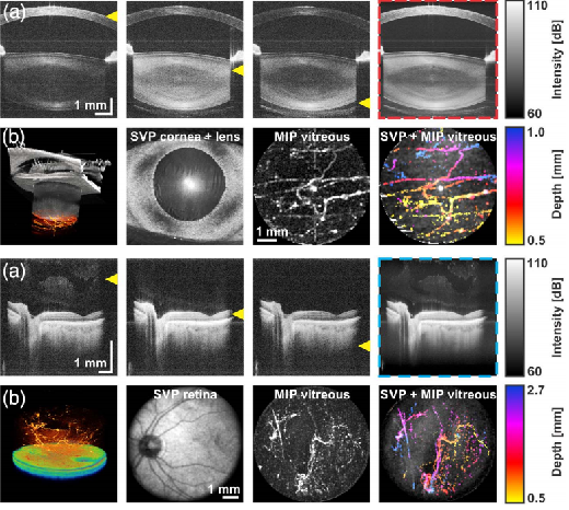
OCT imaging of the anterior segment and retina of the eye using tunable lenses and swept source lighting. (Image: Grulkowski et al.)
Most clinical OCT instruments are limited to imaging depths of two to three millimetres, and with them it is difficult to switch between imaging the front and back portions of the eye due to the eye’s elements that focus light onto the retina.
These challenges are overcome in the researchers’ system by using a tunable lens whose parameters can be controlled dynamically using an electrical current. These varying parameters, unlike the fixed parameters of standard glass and plastic lenses, enable the researchers to focus light in a way that lets them image an entire eye.
‘We incorporated the electrically tunable lens into a custom-made system that represents the latest generation of OCT technology,’ said Grulkowski. ‘We set out to show that we could image both the front and back of the eye without changing instruments. However, we were also able to show that our instrument enhanced the image quality of the OCT images.’
The OCT system also incorporates a newly commercialised swept light source – a laser that continuously changes wavelength very rapidly – that improves the resolution and speed of OCT compared to systems that use other light sources. The researchers also integrated high-speed electronics to achieve the imaging depth necessary to enable whole eye imaging.
By using the new system to image the eyes of seven healthy people, the researchers demonstrated that the resulting images led to measurements that correlated well with those obtained with an ocular biometer, the standard clinical device used today.
The researchers are now working to optimise the instrument for imaging the entire vitreous gel, not just where it interfaces with the lens and retina. The vitreous gel has not been studied intensively and is difficult to image because of its high transparency. The ability to image the entire vitreous could allow OCT to be used to guide procedures that involve the removal of the vitreous gel from the eye, which is sometimes done to repair retinal detachment.
Future developments
Although the laboratory version of the set-up is ready to use, further steps will be taken by the researchers to translate the new system to the clinic.
The researchers are also focused on optimising the scan areas and developing processing tools for the automatic measurement of the dimensions of the eye. These improvements will enable advanced studies of the proposed scan regimes on a group of patients with different types of opacification in the eye.
Related articles