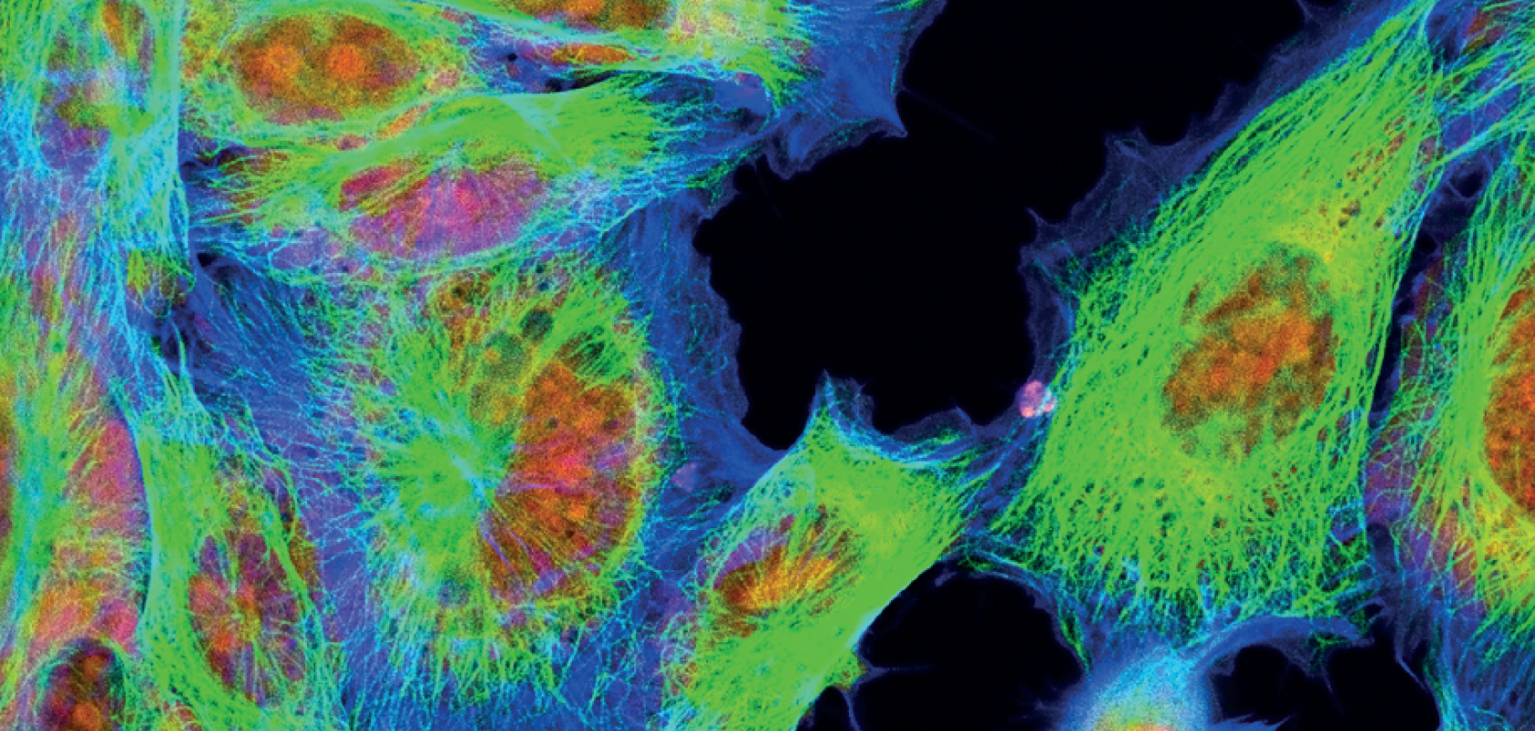
Multiplex microscopy is widely used by researchers, particularly in the biological sciences, to obtain information that will help them gain a greater understanding of biological processes.
The multiplexed fluorescence detection technique can simultaneously detect and quantify multiple targets, such as proteins or nucleic acids in a single sample. This method uses fluorescent dyes or labels that emit distinct signals when excited by different wavelengths of light.
In multiplexed fluorescence detection, multiple fluorescently labelled probes or antibodies are used, each of which targets a specific analyte. Probes are labelled with unique fluorescent dyes, which allows them to be distinguished and detected separately. The sample is then analysed using a specialised instrument, such as a microarray scanner or a flow cytometer, which can detect and measure the fluorescent signals emitted by each dye.
The technique has been used throughout the world for years in applications such as gene expression analysis, protein analysis, pathogen detection, environmental monitoring and drug discovery and development, to name just a few. This is because of the advantages it offers as a cost-effective, accurate, flexible and efficient form of detection.
Multiplexed fluorescence detection: the challenges
However, as demand from biologists and researchers grows to be able to detect more biological targets than ever before, multiplexed fluorescence detection has also encountered its share of challenges – largely due to the risk of spectral crosstalk if more than four or five targets are attempted.
Iain Johnson, Ph.D., Director of Technical Support at Lumencor explains: “In my experience, if you ask a biologist how many samples they need to target, the answer they always provide is ‘more’. If they can identify four, then next they want to identify 40, and then 400 and 4,000. There are billions of different protein molecules in a single cell that produce its activity, and you can only get so far looking at four of them at any one time. So, what happens when four or five is not enough? How do you get to 400, and then 4,000?”
A number of techniques have been developed to help increase the number of targets that can be detected, each with their own advantages and challenges, and each of which requires its own specific chain of component parts that must fit together to guarantee the right results, and each of which relies on the illumination solution as the first step to success. These techniques include the introduction of essential quantum dot nanocrystals with narrow emission spectra, molecular barcode scanning for gene expression analysis, and multiplexed error-robust fluorescence in situ hybridisation (also known as MERFISH).
Looking specifically at MERFISH, the technique works as a single-cell ribonucleic acid (RNA) sequencing method that uses fluorescent probes to detect and quantify multiple RNA targets in individual cells. It can detect hundreds to thousands of genes simultaneously, so it can be a powerful tool for biologists looking to identify more targets.
Continues Johnson: “MERFISH is a state-of-the-art technique where you can actually look at 10,000 targets to some extent. That’s how far things have come. Admittedly, it is a much more complex and expensive technique to implement, but the biological imperative for doing it is so big, the need is so great, that the people who require it are spending large amounts of money and the large amounts of time that it takes to get that answer.”
Multiplexed fluorescence detection: illumination solutions
The MERFISH technique requires the use of multiple colours, which means that selecting the right light source is imperative. A laser source such as Lumecor’s CELESTA laser Light Engine is ideally suited for the task, as Johnson explains: “The light source colour that’s used for this technique is typically an array of five lasers in a box. Because in MERFISH it’s the detecting single molecules present in the cell, it’s a very tiny signal so you have to use a lot of light. The CELESTA produces the amount of light in a very small area that is needed to to actually detect these single molecules, and then the signal that is read out is essentially a barcode that’s used to identify the individual modules. That barcode can be made up of colours, and different colours are used, again for identification of different individuals in the crowd. So you need to be able to switch quickly between the colours, which is achievable.”
Another challenge is that experiments using this technique can take much longer to undertake, due to the incredibly high number of individual identifications. This means that the laser light source has to stay very stable over time for consistent, accurate performance. Johnson elaborates: “If the light source doesn’t remain stable for the longer experiment duration, you could end up in a situation where signals will be crossed, and you won’t be able to tell which target is which. Another big driver is that users want to collect the data quicker, write the same amount of data in less time, so they can get through more experiments in their work. The CELESTA supplies the speed of switching colours, but it also supplies stability over the long timeframe of the experiments.”
With the types of microscopy referred to in this article, there can be a wealth of moving parts, all of which must be correct in order to achieve accurate, repeatable results. There are the microscopes and all of their component parts, cameras, computers, molecular labelling techniques, and of course, the skilled people to carry out the experiments and analyse the data. But before all of this, says Johnson, comes the light.
“The light source is the first piece in this long chain of things that must be connected together to retrieve the data. Light is the first step and Lumencor specialises in supplying that light. It may only be one part of this long, connected chain of elements that are required for multiplexed fluorescence detection and other techniques, but it is the first part, so, in some ways, you could argue it’s the most important part. If you get the first step wrong, then everything after that is likely to also be wrong, and it can be very difficult from which to recover.
“As the starting point of the whole process, it is very important that it be reliable and consistent. That is why it is our focus.”

