A Harvard Research team led by Nobel laureate Eric Betzig has developed a microscope that combines lattice light-sheet microscopy with adaptive optics to capture 3D images and videos of cells inside living organisms in high resolution.
The researchers, from Howard Hughes Medical Institute’s (HHMI) Janelia Research Campus, demonstrated the new technology by generating a series of movies: cancer cells crawling through blood vessels, spinal nerve cells wiring up into circuits, immune cells cruising through a zebrafish’s inner ear, and more.
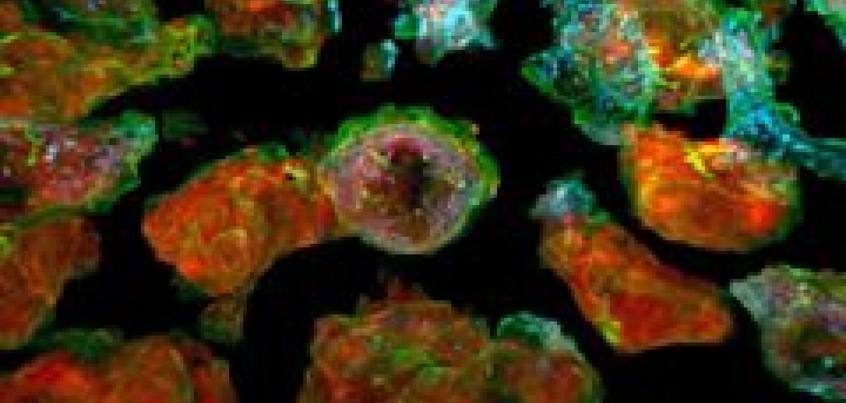
The resolution of the microscope allows subcellular details to be obtained, such as the dynamics of miniscule bubbles known as vesicles, which transport molecular cargo through to the cell.
Credit: Liu et al
‘This is the miracle of being able to see what we have never been able to see before. It’s simply incredible,’ said study co-author Tomas Kirchhausen, HMS professor of cell biology, and the Springer Family Chair of pediatrics and a senior investigator at Boston Children’s Hospital.
‘Every time we’ve done an experiment with this microscope, we’ve observed something novel — and generated new ideas and hypotheses to test,’ Kirchhausen said. ‘It can be used to study almost any problem in a biological system or organism I can think of.’
Visualising living cells in real time inside live organisms is challenging. Cells of interest are surrounded by tissues and other biological structures that scramble light coming from and returning to a microscope objective, which blurs and obscures important details. Light powerful enough to penetrate biological structures and yield a crisper view of cells, on the other hand, can damage tissues.
Guide star
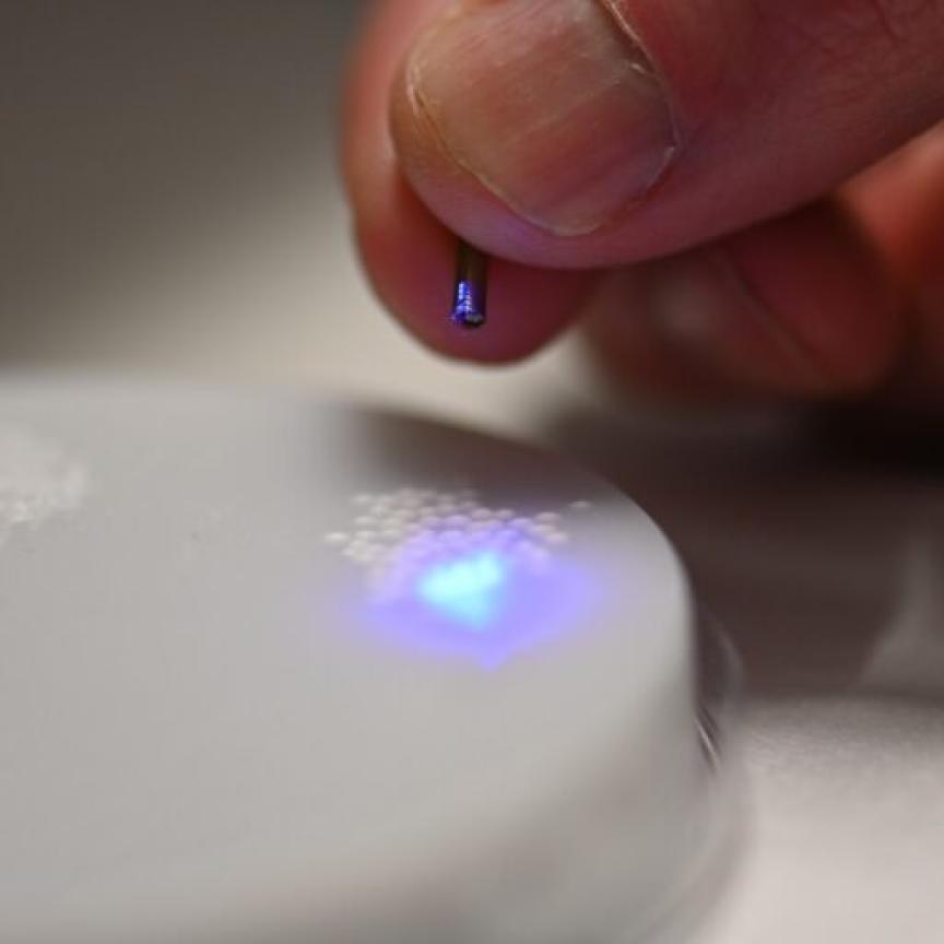
To address these challenges, Betzig and colleagues combined two technologies: lattice light-sheet microscopy, which Betzig developed in the early 2010s, and adaptive optics, a technique borrowed from astronomy.
Lattice light-sheet microscopy uses rapid and repeated sweeps of an ultrathin sheet of light, which avoids the bleaching or damage associated with traditional focused beams of light. It generates high-resolution 2D images of living cells as they carry out their functions, and by combining series of these images over time, scientists can create 3D movies.
To unscramble the light sheet as it passes through tissues and other structures, the research team used adaptive optics.
Astronomers often use adaptive optics to counteract the effects of atmospheric turbulence. The process uses a high-power laser, aimed at the small region of the sky they want to image, which serves as a ‘guide star.’ As the laser passes through the atmosphere, optical aberrations that distort its path are revealed and corrected by the adaptive optics such as deformable mirrors and light modulators.
Betzig and colleagues applied this principle to the microscopic world, using a two-photon laser to create an adaptive optics system that maintains the thin illumination of a lattice light sheet as it penetrates an organism to generate distortion-free images of their target.
The team validated the new adaptive optics/lattice light-sheet microscope on a variety of biological samples, carrying out much of the work through the laboratories of Kirchhausen and Sean Megason, HMS associate professor of systems biology.
To make sense of the data they generated, the team developed software and computational and visualisation workflows, spearheaded by study co-lead authors Gokul Upadhyayula, HMS instructor in pediatrics at Boston Children’s and research associate in the Kirchhausen lab, and Tsung-Li Liu, formerly a research scientist in the Betzig lab at HHMI.
‘For the types of data we generated, there’s no one commercial software that we can use to create interpretable movies and extract biologically meaningful information, so we built the necessary tools,’ Upadhyayula said. ‘This allowed us to understand what we acquired and visualise the data in meaningful ways, including, in the near future, fully interactive 3D movies.’
The team captured movies of the behavior of organelles as they remodel themselves inside cells during cell division, and even visualised in real-time and at near-molecular detail the process of clathrin-mediated endocytosis, which cells use to capture materials from their exterior environment.
‘I work on understanding how cells "eat" using machinery based on vesicular carriers, and all my life I’ve dreamed of seeing this in a live organism,’ Kirchhausen said. ‘We have finally achieved this.’
The complexity of the 3-D multicellular environment can be overwhelming, Betzig said, but the clarity of the imaging allows the researchers to computationally ‘explode’ apart the individual cells in tissue to focus on the dynamics within any particular one.
‘It’s like ‘Star Trek.’ It’s the age of exploration again,’ Upadhyayula said. ‘We don’t even know what questions to ask yet because we’ve never even seen some of these biologies at this level of detail.’
All this detail is hard to see without adaptive optics, Betzig said. ‘It’s just too damn fuzzy.’ In his view, adaptive optics is one of the most important areas in microscopy research today, and the lattice light-sheet microscope, which excels at 3D live imaging, is the perfect platform to showcase its power.
The next step is making the technology affordable and user-friendly. The current microscope takes up a 10-foot-long table. In collaboration with Kirchhausen and Upadhyayula, Betzig’s team is working on a next-generation version that should fit on a small desk at a cost within the reach of individual labs.
The first such instrument will go to Janelia’s Advanced Imaging Centre, where scientists from around the world can apply to use it. A second instrument built at the same time will be located in the Kirchhausen laboratory at the HMS Quad in Boston. Plans to build the instrument will also be made available.