Revealing cellular mysteries with ultrafast lasers

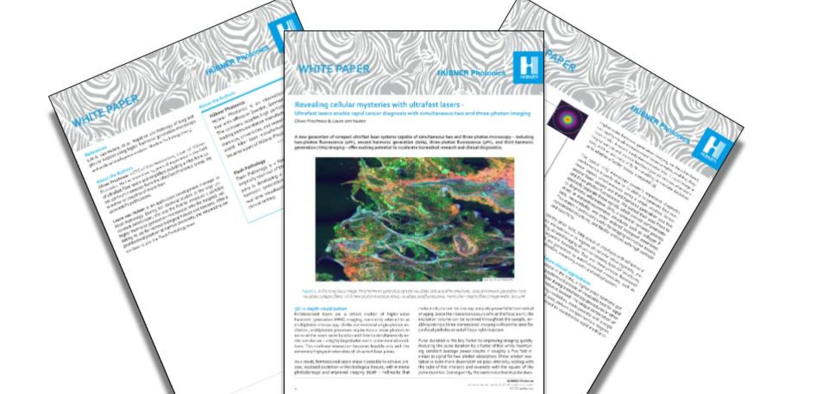
Revealing cellular mysteries with ultrafast lasers - A Hübner Photonics White Paper
The ability to visualise cellular structures and tissue architecture in three dimensions, without staining or sample preparation, has long been a goal of biomedical research. Traditional microscopy techniques require time-consuming sample slicing, staining, and processing, creating delays that can prove critical in clinical settings. Meanwhile, the wait for histopathological analysis can take several days, potentially requiring patients to undergo repeated procedures when initial biopsies prove inconclusive.
A new era of multiphoton microscopy
This White Paper introduces a breakthrough: compact ultrafast laser systems capable of simultaneous two- and three-photon microscopy. These advanced femtosecond lasers enable higher harmonic generation (HHG) imaging, including two-photon fluorescence (2PF), second harmonic generation (SHG), three-photon fluorescence (3PF), and third harmonic generation (THG), all from a single laser source. The result is label-free, high-resolution imaging of fresh, unprocessed biological specimens with unprecedented speed and accuracy.
Who should read this White Paper?
This resource is designed for:
- Biomedical researchers seeking advanced imaging capabilities for cellular and tissue analysis
- Clinical diagnosticians looking to accelerate intraoperative decision making
- Pathologists interested in rapid tissue analysis techniques
- Medical device developers working on next-generation diagnostic systems
- Academic researchers exploring multiphoton microscopy applications
- Healthcare professionals involved in bronchoscopy and tissue biopsy procedures
- Anyone interested in how femtosecond laser technology is revolutionising medical diagnostics
What you'll discover
Inside this detailed White Paper, you'll explore:
- The science behind the innovation: Understanding how femtosecond lasers enable multiphoton processes through extremely high peak intensities, creating precise, localised excitation within biological tissues with minimal photodamage
- Technical advances: Why pulse duration is critical to imaging quality, and how sub-40 femtosecond pulses with ultrabroadband optical spectra deliver significantly higher peak power and unmatched imaging contrast
- Simultaneous multi-modal imaging: How a single laser source generates four complementary nonlinear signals: SHG revealing collagen fibres and connective tissues, THG visualising cell membranes and organelle boundaries, plus two- and three-photon fluorescence highlighting autofluorescent molecules
- Clinical applications: Real-world examples including rapid cancer diagnosis during bronchoscopy, with demonstrated accuracy of 87% and diagnostic feedback delivered just six minutes after tissue excision
- Future possibilities: The potential for in situ tissue analysis across various surgical procedures, eliminating the need for repeat biopsies and accelerating patient care pathways
Accelerate your research and clinical capabilities
Download this White Paper to discover how the VALO Femtosecond Series and similar advanced ultrafast laser systems are unlocking new possibilities in biomedical imaging, from fundamental research to revolutionary clinical diagnostics that could transform patient outcomes.

