How Oxxius MixxWave laser combiners power UCL's cancer research mesoSPIM for lightsheet brain imaging
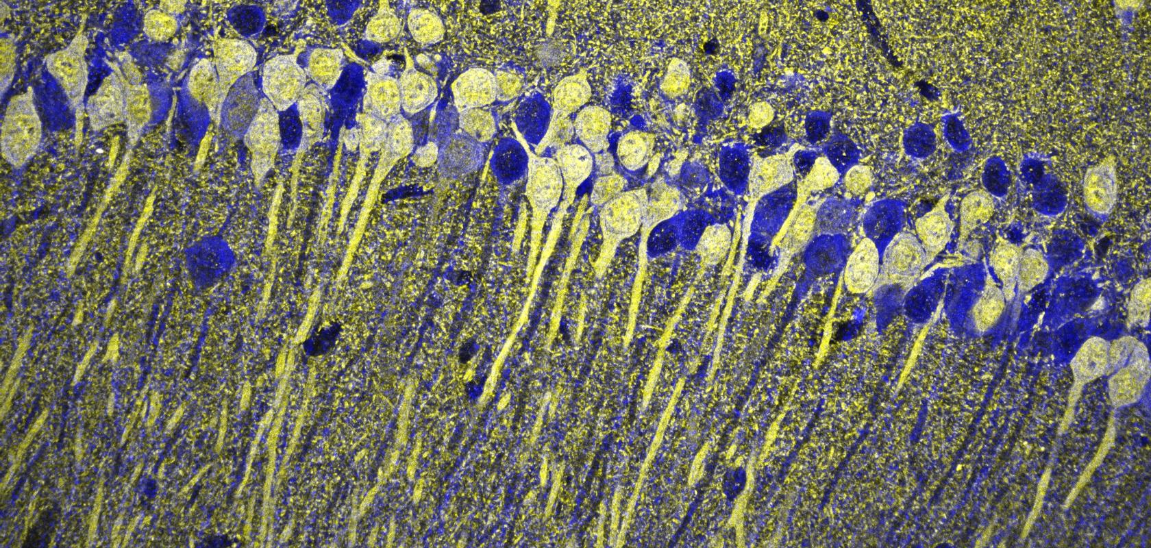
Image of a section of mouse hippocampus (Credit: Alexandros A Lavdas/Shutterstock.com)
Understanding how cancer invades, metastasises and resists treatment requires seeing tumours as they truly exist: complex, three-dimensional cellular ecosystems with heterogeneous spatial gradients in cell density, oxygenation and immune infiltration.
Traditional microscopy techniques can struggle with this challenge. Confocal systems are too slow for large cleared samples and cause photobleaching. Commercial lightsheet platforms exist but remain expensive and closed to modification. Sectioning destroys the morphological context that reveals invasion corridors and micro-metastases.
Lightsheet microscopy for cancer research
Lightsheet microscopy offers a transformative solution, illuminating only the imaging plane to acquire large volumes quickly with minimal photodamage. When combined with tissue clearing methods and multicolour fluorescence excitation, lightsheet systems can image intact tumours and whole brains at subcellular resolution whilst preserving biological context across centimetre-scale specimens.
This White Paper examines how University College London's Cancer Institute has deployed an open-source mesoSPIM lightsheet microscope powered by Oxxius MixxWave laser combiners to advance brain cancer research, demonstrating why compact, stable multicolour illumination has become infrastructure for cancer discovery.
Who should read this White Paper?
This resource is essential for:
- Cancer researchers investigating tumour micro-environments, invasion patterns and treatment responses in whole-organ contexts
- Imaging facility managers evaluating scalable lightsheet platforms and flexible laser sources for translational research
- Neuroscientists studying brain tumours, particularly gliomas that infiltrate neural structures and co-opt neuronal circuits
- Microscopy specialists seeking practical insights into integrating modular laser combiners with open-source imaging systems
What you'll discover
The White Paper details both the technical capabilities and practical workflow advantages of combining Oxxius MixxWave laser combiners with the mesoSPIM platform, showing how this pairing enables cancer studies that would be impossible with conventional microscopy.
Key insights include:
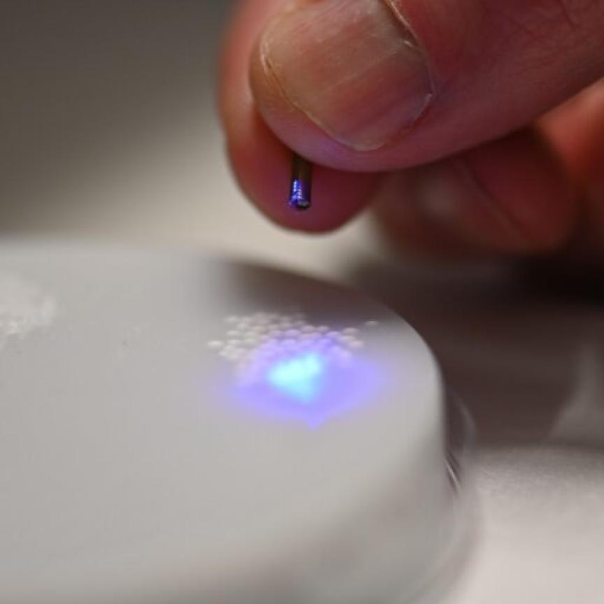
MixxWave combiner architecture: how modular L4Cc/L6Cc platforms consolidate four to six wavelength channels (cyan through far-red, extending to 1064 nm) into one compact unit with fibre-coupled outputs, direct modulation or AOM control, and excellent power stability
mesoSPIM integration advantages: fibre delivery that eliminates beam walk and simplifies daily start-up in busy facilities, synchronised multi-channel acquisition for dynamic timelapses, and scalability that allows wavelength additions without redesigning the entire light source
Cancer research applications: whole-brain tumour mapping in mouse models revealing invasion corridors missed by conventional sectioning, 3D vascular and immune micro-environment analysis quantifying vessel density and infiltration patterns, and high-throughput drug screening with volumetric readouts
Unique NIR capabilities: single-mode fibre delivery extended from the typical 660 nm limit to 1064 nm, enabling excitation of NIR fluorophores like Alexa Fluor 680 for deeper sample penetration and additional discrimination levels
The White Paper includes specific use cases from UCL's installation, where researchers visualise clonal cancer cell populations in cleared mouse brains, trace neural connectivity alterations caused by gliomas, and correlate micro-environment features with therapeutic response across entire organs.
Why this matters for translational research
Brain tumours present distinctive imaging challenges: infiltrative growth that follows white matter tracts, perivascular spread creating distant micro-metastases, and delicate neural structures disrupted by mechanical sectioning. Small 2D sections cannot capture these spatial relationships or reveal the heterogeneous gradients that define tumour biology.
By combining tissue clearing protocols with mesoSPIM's millimetre-to-centimetre field of view and Oxxius combiners' multicolour excitation, UCL's platform delivers the throughput, spatial context and quantitative data required for computational pipelines performing cell segmentation, vascular network analysis and machine-learning-driven phenotyping across experimental conditions.
This combination transforms illumination from a technical specification into research infrastructure, enabling biologically faithful, high-throughput 3D imaging that reveals how tumours actually behave within their organ environments.