Using the full spectrum for Raman: from UV to NIR
Wavelength is key
The physical basis of the technique offers huge flexibility and advantages in comparison with its sister technique, infrared spectroscopy, but at the same time presents a key challenge: the excitation needs to be highly (i) monochromatic (the Raman bands have the same shape as the light source) (ii) collimated and (iii) intense (due to the low probability of inelastic scattering, < 1 in 106 photons). Hence, it is the advent of lasers that has brought Raman spectroscopy – literarily –into the field.

Figure 1: Near-IR dispersive Raman Spectroscopy. Comparison of the spectrum of a solid sample of an organic flurophore at 785 (red) with that obtained at 1064 nm (blue). Excitation with a Cobolt RumbaTM 1064 nm (100 mW at sample), detection 4 by 5 s exposure, Andor idus163 spectrograph with an iDus-InGaAs detector.
In selecting the correct laser, the wavelength is important and depends on the application. For example, fluorescence is a usually much stronger than the Raman scattering signal but unlike the fluorescence, the Raman scattering signal is observed even when exciting at wavelengths outside the absorption spectrum. Figure 1 illustrates this by showing that the fluorescence that swamps the Raman spectrum of a boron based fluorophore when the Raman spectrum is recorded at 785 nm (where absorption/fluorescence competes with the Raman process) is completely absent in the spectrum obtained at 1064 nm. Thus highlighting the importance of selecting the right excitation wavelength.
DPSS lasers for Raman
Until recently, ion gas lasers (e.g. Ar, He, HeCd and Kr) have been the first choice for Raman spectroscopy. However, the ever increasing number of wavelengths available with continuous wave diode pumped solid state (DPSS) lasers together with the high average powers (> 1 W) and compact footprint means that multi-wavelength Raman spectroscopy can be implemented as a turn key low maintenance solution in any laboratory as well as in field portable applications. For example, Cobolt DPSS lasers qualify as excellent Raman excitation sources, thanks to their extremely narrow linewidth (<1MHz), excellent wavelength stability and high level of spectral purity (>-60dB).

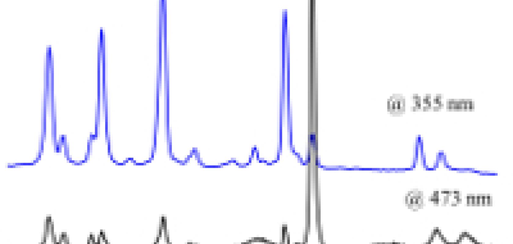
Figure 2: Raman spectra of a copper (II) complex (1 mM in water) obtained using 355 nm (Cobolt ZoukTM, 10 mW) and at 473 nm (Cobolt BluesTM 50 mW). 355 nm is close to the electronic absorption of the copper (II) complex and hence the spectrum is much more intense than would be expected to be due to resonance enhancement effect.
Raman scattering is inherently weak – or is it?
Although it is true that the number of inelastically scattered photons is vanishing small, this does not mean that Raman spectroscopy cannot be used to detect compounds at low concentrations (e.g. < 1 mM) or that spectra cannot be obtained rapidly. When the laser used is close to or the same wavelength as a compound absorbs light (i.e. is in resonance) then the Raman signal from that compound can be enhanced by up to 104 times. An example of the resonance Raman effect is shown in Figure 2; by choosing 355 nm rather than 473 nm laser excitation the observed Raman signals are much stronger thus providing improved resolution.
Conclusion
With the current availability of high performance DPSS lasers offering a broad range of wavelengths from the UV to the NIR, the applications for Raman spectroscopy are only just beginning to be fully explored.
References
1. P. Dijkstra, Davide Angelone, E. Taknishnikh, H. J. Wörtche, E. Otten, W. R. Browne, Dalton, 2014, 10.1039/C4DT01393J
2. W. R. Browne, J. J. McGarvey, Coord. Chem. Rev., 2007, 251, 454-473.
Wavelength is key
The physical basis of the technique offers huge flexibility and advantages in comparison with its sister technique, infrared spectroscopy, but at the same time presents a key challenge: the excitation needs to be highly (i) monochromatic (the Raman bands have the same shape as the light source) (ii) collimated and (iii) intense (due to the low probability of inelastic scattering, < 1 in 106 photons). Hence, it is the advent of lasers that has brought Raman spectroscopy – literarily –into the field.
Figure 1: Near-IR dispersive Raman Spectroscopy. Comparison of the spectrum of a solid sample of an organic flurophore at 785 (red) with that obtained at 1064 nm (blue). Excitation with a Cobolt RumbaTM 1064 nm (100 mW at sample), detection 4 by 5 s exposure, Andor idus163 spectrograph with an iDus-InGaAs detector.
In selecting the correct laser, the wavelength is important and depends on the application. For example, fluorescence is a usually much stronger than the Raman scattering signal but unlike the fluorescence, the Raman scattering signal is observed even when exciting at wavelengths outside the absorption spectrum. Figure 1 illustrates this by showing that the fluorescence that swamps the Raman spectrum of a boron based fluorophore when the Raman spectrum is recorded at 785 nm (where absorption/fluorescence competes with the Raman process) is completely absent in the spectrum obtained at 1064 nm. Thus highlighting the importance of selecting the right excitation wavelength.
DPSS lasers for Raman
Until recently, ion gas lasers (e.g. Ar, He, HeCd and Kr) have been the first choice for Raman spectroscopy. However, the ever increasing number of wavelengths available with continuous wave diode pumped solid state (DPSS) lasers together with the high average powers (> 1 W) and compact footprint means that multi-wavelength Raman spectroscopy can be implemented as a turn key low maintenance solution in any laboratory as well as in field portable applications. For example, Cobolt DPSS lasers qualify as excellent Raman excitation sources, thanks to their extremely narrow linewidth (<1MHz), excellent wavelength stability and high level of spectral purity (>-60dB).
Figure 2: Raman spectra of a copper (II) complex (1 mM in water) obtained using 355 nm (Cobolt ZoukTM, 10 mW) and at 473 nm (Cobolt BluesTM 50 mW). 355 nm is close to the electronic absorption of the copper (II) complex and hence the spectrum is much more intense than would be expected to be due to resonance enhancement effect.
Raman scattering is inherently weak – or is it?
Although it is true that the number of inelastically scattered photons is vanishing small, this does not mean that Raman spectroscopy cannot be used to detect compounds at low concentrations (e.g. < 1 mM) or that spectra cannot be obtained rapidly. When the laser used is close to or the same wavelength as a compound absorbs light (i.e. is in resonance) then the Raman signal from that compound can be enhanced by up to 104 times. An example of the resonance Raman effect is shown in Figure 2; by choosing 355 nm rather than 473 nm laser excitation the observed Raman signals are much stronger thus providing improved resolution.
Conclusion
With the current availability of high performance DPSS lasers offering a broad range of wavelengths from the UV to the NIR, the applications for Raman spectroscopy are only just beginning to be fully explored.
References
1. P. Dijkstra, Davide Angelone, E. Taknishnikh, H. J. Wörtche, E. Otten, W. R. Browne, Dalton, 2014, 10.1039/C4DT01393J
2. W. R. Browne, J. J. McGarvey, Coord. Chem. Rev., 2007, 251, 454-473.